. 2009 Nov;29(22):6006-17.
doi: 10.1128/MCB.01921-08. Epub 2009 Sep 14.

Nuclear factor I-C links platelet-derived growth factor and transforming growth factor beta1 signaling to skin wound healing progression

Affiliations

Nuclear factor I-C links platelet-derived growth factor and transforming growth factor beta1 signaling to skin wound healing progression

Genta Plasari et al. Mol Cell Biol. 2009 Nov.

Abstract

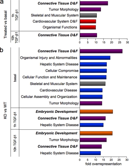
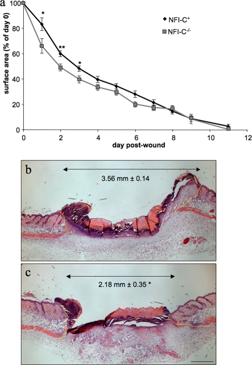
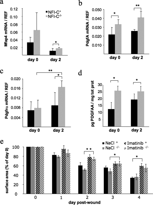
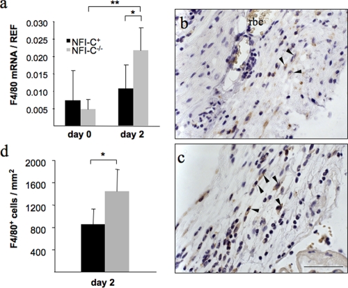
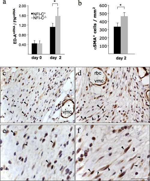
Transforming growth factor beta (TGF-beta) and platelet-derived growth factor A (PDGFAlpha) play a central role in tissue morphogenesis and repair, but their interplay remain poorly understood. The nuclear factor I C (NFI-C) transcription factor has been implicated in TGF-beta signaling, extracellular matrix deposition, and skin appendage pathologies, but a potential role in skin morphogenesis or healing had not been assessed. To evaluate this possibility, we performed a global gene expression analysis in NFI-C(-/-) and wild-type embryonic primary murine fibroblasts. This indicated that NFI-C acts mostly to repress gene expression in response to TGF-beta1. Misregulated genes were prominently overrepresented by regulators of connective tissue inflammation and repair. In vivo skin healing revealed a faster inflammatory stage and wound closure in NFI-C(-/-) mice. Expression of PDGFA and PDGF-receptor alpha were increased in wounds of NFI-C(-/-) mice, explaining the early recruitment of macrophages and fibroblasts. Differentiation of fibroblasts to contractile myofibroblasts was also elevated, providing a rationale for faster wound closure. Taken together with the role of TGF-beta in myofibroblast differentiation, our results imply a central role of NFI-C in the interplay of the two signaling pathways and in regulation of the progression of tissue regeneration.


Figures

FIG. 1.
FIG. 1.
Specific biological functions ascribed to TGF-β1 and NFI-C potential target genes. The percentage of misregulated genes assigned to each biological category was normalized to that of all genes on the microarray pertaining to the considered category. Illustrated are the functions that were overrepresented more than 10-fold in the early (1 h) and late (10 h) TGF-β1 response (a) and more than 13-fold in the comparisons of NFI-C-null versus wild-type fibroblasts (b). D&F, “development and function”; KO, knockout; WT, wild type. Red and blue bars indicate function targeted by only TGF-β1 or only NFI-C, respectively, while the violet ones depict functions potentially regulated by both TGF-β1 and NFI-C. Gray bars indicate functions targeted by TGF-β1 and NFI-C but not by their combination, while orange bars implicate both regulatory molecules together but not either one alone.
FIG. 2.
FIG. 2.
Acceleration of skin wound healing in NFI-C−/− mice. (a) Kinetics of wound closure in wild-type and NFI-C knockout mice. The surfaces of wounds were recorded daily and plotted as percentage of the initial wound area at day 0, and they are represented as the mean of the surface of 12 wounds ± the standard error of the mean (SEM). (b and c) Hematoxylin and eosin staining of wild-type (b) and NFI-C-null (c) wounds at day 2. Yellow dashed lines border the injured tissue. The distance between healthy dermises (double arrow) was measured in sections of four wild-type and four knockout mice, and the average value ± SEM is indicated in each panel. *, P ≤ 0.05; **, P ≤ 0.01. Bar, 500 μm.
FIG. 3.
FIG. 3.
Expression of PDGF signaling intermediates and Mfap4 in the wounded skin of NFI-C-null and wild-type mice. RT-qPCR assays were performed on mRNAs extracted from normal (day 0) or wounded (day 2) skin samples to evaluate gene expression of Mfap4 (a), PdgfA (b), and Pdgfrα (c). The level of expression was normalized to endogenous reference genes as described in Materials and Methods. Values represent the means of five mice ± the standard deviation. (d) Quantification of PDGFA in skin protein extracts was determined by ELISA, and values were normalized to the total protein content. Values represent the means of four mice ± the standard error of the means (SEM). (e) Quantification of wound closure after injury in wild-type and knockout mice treated with a saline solution or with imatinib mesylate (Gleevec). Values represent the means of three mice ± SEM. *, P ≤ 0.05; **, P ≤ 0.01.
FIG. 4.
FIG. 4.
Increased infiltration of macrophages in the wounds of NFI-C-deficient mice. (a) RT-qPCR assays of mRNA extracted from skin samples were performed to evaluate the expression of a macrophage marker, F4/80 (EGF-like module and mucin-like, hormone receptor-like sequence), in NFI-C−/− mice and control mice at day 0 and day 2 postwounding. The level of expression was normalized to endogenous reference genes as described in Materials and Methods. Values represent the means of five mice ± the standard deviation. F4/80 protein levels in injured skin was assessed by the immunostaining of granulation tissues at day 2 postwounding in wild-type (b) and knockout (c) animals. The arrowheads indicate all the F4/80-positive cells in the subsected fields. Tissues were counterstained with hematoxylin. Staining of red blood cells (rbc) is due to the endogenous presence of peroxidase. Bar, 50 μm. (d) F4/80-positive cells were quantified in five control (black column) and five knockout (gray column) mice (mean ± the standard error of the mean) and normalized to the wounded area as described in Materials and Methods. *, P ≤ 0.05.
FIG. 5.
FIG. 5.
Early myofibroblast differentiation accompanies enhanced wound healing in knockout animals. The expression levels of two genes involved in the myofibroblastic phenotype are shown as follows. (a) The spliced variant of fibronectin (ED-A FN) was assayed by RT-qPCR performed on mRNA extracted from skin samples of NFI-C−/− and control mice at day 0 and day 2 postwounding. The level of expression was normalized to the total fibronectin mRNA as described in Materials and Methods. Values represent the means of five mice ± the standard deviation. (b) αSMA-positive cells were labeled and quantified in five control mice (black column) and five knockout mice (gray column) (means ± standard error of the mean) and normalized to the wounded area as described in Materials and Methods. *, P ≤ 0.05. Immunostaining of the myofibroblastic marker αSMA in the granulation tissue of wild-type (c) and knockout (d) animals (panels e and f are enlargements of panels c and d, respectively) at day 2 postwounding. The arrowheads indicate the αSMA-positive cells in a subset of the fields. Vascular muscle cells (vmc) and red blood cells (rbc) are indicated to show similar staining of αSMA vessels in normal or knockout mice. Tissues were counterstained with hematoxylin. Bars, 25 μm.

References

    1. Alevizopoulos, A., Y. Dusserre, M. Tsai-Pflugfelder, T. von der Weid, W. Wahli, and N. Mermod. 1995. A proline-rich TGF-beta-responsive transcriptional activator interacts with histone H3. Genes Dev. 9:3051-3066. - PubMed
    1. Alonso, C. R., C. G. Pesce, and A. R. Kornblihtt. 1996. The CCAAT-binding proteins CP1 and NF-I cooperate with ATF-2 in the transcription of the fibronectin gene. J. Biol. Chem. 271:22271-22279. - PubMed
    1. Arici, A., P. C. MacDonald, and M. L. Casey. 1996. Modulation of the levels of transforming growth factor beta messenger ribonucleic acids in human endometrial stromal cells. Biol. Reprod. 54:463-469. - PubMed
    1. Baker, J., J. P. Liu, E. J. Robertson, and A. Efstratiadis. 1993. Role of insulin-like growth factors in embryonic and postnatal growth. Cell 75:73-82. - PubMed
    1. Balza, E., L. Borsi, G. Allemanni, and L. Zardi. 1988. Transforming growth factor beta regulates the levels of different fibronectin isoforms in normal human cultured fibroblasts. FEBS Lett. 228:42-44. - PubMed

Publication types

MeSH terms

Substances