. 2009 Sep 15;120(11 Suppl):S230-7.
doi: 10.1161/CIRCULATIONAHA.108.841155.

Novel minicircle vector for gene therapy in murine myocardial infarction

Affiliations

Novel minicircle vector for gene therapy in murine myocardial infarction

Mei Huang et al. Circulation. .

Abstract

Background: Conventional plasmids for gene therapy produce low-level and short-term gene expression. In this study, we develop a novel nonviral vector that robustly and persistently expresses the hypoxia-inducible factor-1 alpha (HIF-1alpha) therapeutic gene in the heart, leading to functional benefits after myocardial infarction.

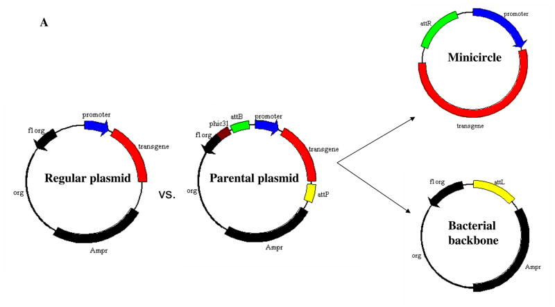
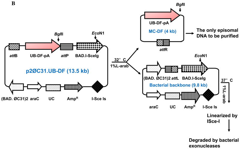
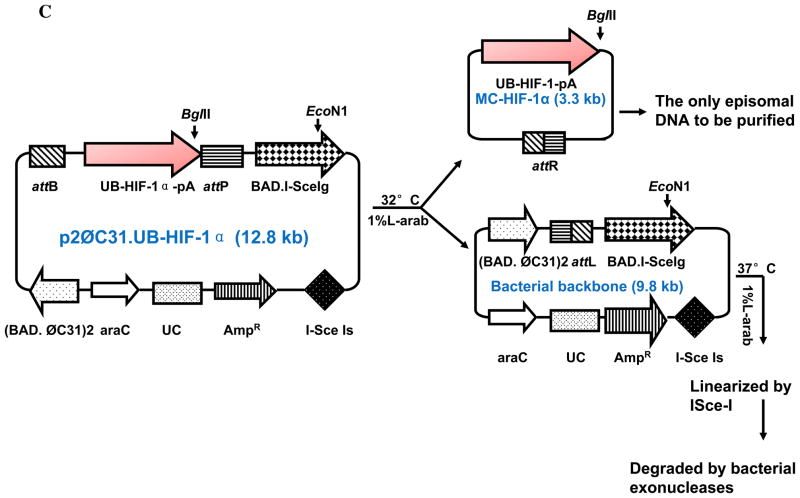
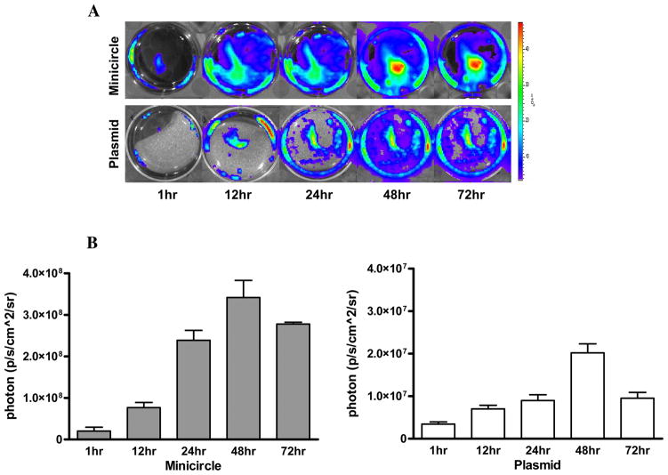
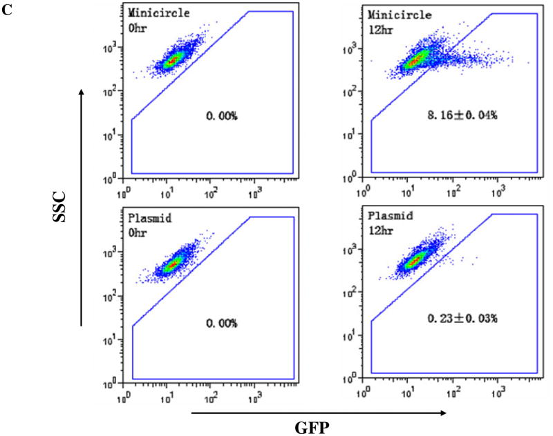
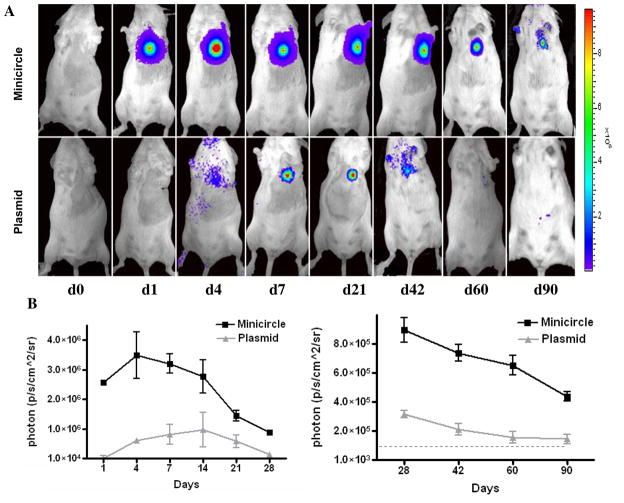
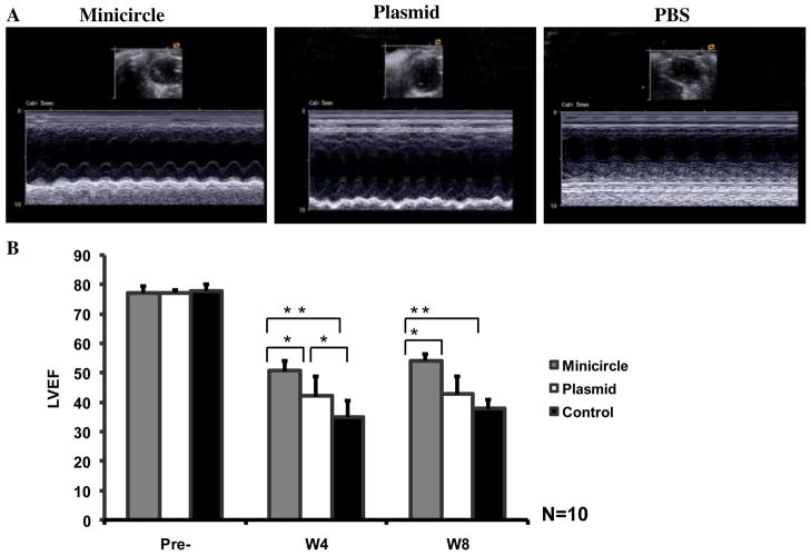
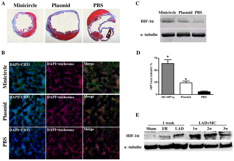
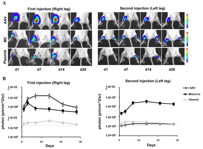
Methods and results: We first created minicircles (MC) carrying double-fusion reporter gene consisting of firefly luciferase and enhanced green fluorescent protein (Fluc-eGFP) for noninvasive measurement of transfection efficiency. Mouse C2C12 myoblasts and normal FVB/N mice were used for in vitro and in vivo confirmation, respectively. Bioluminescence imaging showed stable MC gene expression in the heart for >12 weeks and the activity level was 5.6+/-1.2-fold stronger than regular plasmid at day 4 (P<0.01). Next, we created MC carrying HIF-1alpha (MC-HIF-1alpha) therapeutic gene for treatment of myocardial infarction. Adult FVB/N mice underwent left anterior descending ligation and were injected intramyocardially with: (1) MC-HIF-1alpha; (2) regular plasmid carrying HIF-1alpha (PL-HIF-1alpha) as positive control; and (3) PBS as negative control (n=10/group). Echocardiographic study showed a significantly greater improvement of left ventricular ejection fraction in the MC group (51.3%+/-3.6%) compared to regular plasmid group (42.3%+/-4.1%) and saline group (30.5%+/-2.8%) at week 4 (P<0.05 for both). Histology demonstrated increased neoangiogenesis in both treatment groups. Finally, Western blot showed MC express >50% higher HIF-1alpha level than regular plasmid.

Conclusions: Taken together, this is the first study to our knowledge to demonstrate that MC can significantly improve transfection efficiency, duration of transgene expression, and cardiac contractility. Given the serious drawbacks associated with most viral vectors, we believe this novel nonviral vector can be of great value for cardiac gene therapy protocols.


Figures

Figure 1
Figure 1. Schema of the non-viral minicircle plasmid
(A) Minicircles are the product of site-specific recombination between the attB and attP sites driven by bacteriophage ΦC31 integrase. (B) Schema of the production process for minicircles carrying Fluc-eGFP double fusion reporter gene. By adding 1%-L-arabinose to the bacterial culture media, the att sites of p2øC31.UB-DF were recombined to generate the minicircle DNA. The remaining circular bacterial backbone plasmids were linearized by I-SceI homing endonuclease and were removed by bacterial exonucleases at 37°C. (C) Schema of the production process for minicircles carrying HIF-1α therapeutic gene. In all three schemas, the end result is two circular DNAs: one is the minicircle (MC), which contains the therapeutic gene cassette and the right hybrid sequence (attR), and the other is the bacterial backbone, which contains the origin of replication, the antibiotic marker, and the left hybrid sequence (attL).
Figure 2
Figure 2. Comparison of minicircles vs. regular plasmids in vitro.
(A) The DF consists of Fluc and eGFP linked by a 5-amino acid linker (GSHGD). In vitro BLI shows that Fluc signals are significantly higher in C2C12 cells transfected with minicircles compared to plasmids at all time points. (B) Quantitation of Fluc indicates that minicircles are 5.5±1.7 (at 12 hr) and 8.1±2.8-fold (at 48 hr) higher than regular plasmid. Note the difference in Y-axis bars between the two plots. (C) eGFP expression through FACS at 12 hr coincides with the bioluminescence imaging results.
Figure 3
Figure 3. Comparison of minicircles vs. regular plasmids in vivo.
(A) Both MC-DF and PL-DF were injected into normal murine hearts. Mice injected with minicircles (top row) showed more robust Fluc signals compared to mice injected with regular plasmid (bottom row). Transgene expression was detectable at day 1, peaked at week 1–2, and lasted for >90 days. (B) Detailed quantitative analysis of Fluc bioluminescence signals from days 1–28 (left) and days 28–90 (right). Note the difference in Y-axis scale bars (as p/sec/cm2/sr) between the two plots. Background bioluminescence signal is denoted by the dashed line (1.33×10⁴ p/sec/cm2/sr).
Figure 4
Figure 4. Evaluation of cardiac function following minicircle vs. plasmid mediated HIF-1α therapy
(A) Representative echocardiogram (M-mode) of mice with LAD ligation following injection of minicircles (left), regular plasmid (middle), or PBS (right) as control group at week 8. (B) Quantitative analysis of left ventricular ejection fraction (LVEF) among the three groups. Compared to saline injection, animals injected with MC-HIF-1α had significant improvements in LVEF at week 4 and week 8. Animals injected with regular plasmids had significant improvement in LVEF at week 4 but not by week 8.
Figure 5
Figure 5. Confirmation of HIF-1α overexpression in postmortem explanted hearts
(A) Representative H&E staining shows preservation of thicker heart wall mass after MC-HIF-1α treatment compared to PL- HIF-1α or PBS injections at week 8. (B) Immunofluorescence staining of CD31 endothelial marker (green) indicates increased small vessels in the myocardium following MC-HIF-1α and PL-HIF-1α therapy compared to PBS control. Cardiomyocyte staining is identified by trichrome (red; 100x magnification) Nuclear staining is identified by DAPI (blue; 100x magnification). (C-D) Representative Western blots and quantitative densitometric analysis of explanted hearts injected with MC-HIF-1α, PL-HIF-1α or PBS control at day 14. Significant upregulation of HIF-1α can be seen in the minicircle group. (E) Western blot shows higher activation of endogenous HIF-1α by LAD ligation compared to ischemia-reperfusion. Following delivery of MC-based gene therapy, HIF-1α levels are most robust at week 1 and decreases subsequently, coinciding with similar pattern of Fluc imaging signal decay seen in Figure 3A. Sham: open thoracotomy only; I/R, ischemia/reperfusion; LAD: LAD ligation.
Figure 6
Figure 6. Comparison of viral vs. non-viral mediated gene expression
(A) Representative BLI images of animals injected with AAV, minicircle, and regular plasmid in the right leg (first injection) followed by the left leg 28 days later (second injection). As expected, AAV expression is more robust compared to MC and plasmids initially. However, following repeat injection, AAV expression is not detected in the contralateral leg due to host mediated humoral immune respone. Color scale bar values are expressed as photons per second per square centimeter per steradian (p/s/cm2/sr). (B) Graphical representation of longitudinal BLI after first and second injections in all three groups. Note that day 28 of second injection in left leg would represent day 56 of first injection in right leg in the same animal.

References

    1. Lyon AR, Sato M, Hajjar RJ, Samulski RJ, Harding SE. Gene therapy: targeting the myocardium. Heart. 2008;94:89–99. - PubMed
    1. Marshall E. Gene therapy death prompts review of adenovirus vector. Science. 1999;286:2244–2245. - PubMed
    1. Edelstein ML, Abedi MR, Wixon J. Gene therapy clinical trials worldwide to 2007--an update. The Journal of Gene Medicine. 2007;9:833–842. - PubMed
    1. Jechlinger W. Optimization and delivery of plasmid DNA for vaccination. Expert Review of Vaccines. 2006;5:803–825. - PubMed
    1. Jechlinger W, Azimpour Tabrizi C, Lubitz W, Mayrhofer P. Minicircle DNA immobilized in bacterial ghosts: in vivo production of safe non-viral DNA delivery vehicles. Journal of Molecular Microbiology and Biotechnology. 2004;8:222–231. - PubMed

Publication types

Substances