

Phosphodiesterase-5 inhibitor, tadalafil, protects against myocardial ischemia/reperfusion through protein-kinase g-dependent generation of hydrogen sulfide
- PMID: 19752383
- PMCID: PMC4230451
- DOI: 10.1161/CIRCULATIONAHA.108.843979
Phosphodiesterase-5 inhibitor, tadalafil, protects against myocardial ischemia/reperfusion through protein-kinase g-dependent generation of hydrogen sulfide
Erratum in
- Circulation. 2009 Oct 13;120(15):e139
Abstract
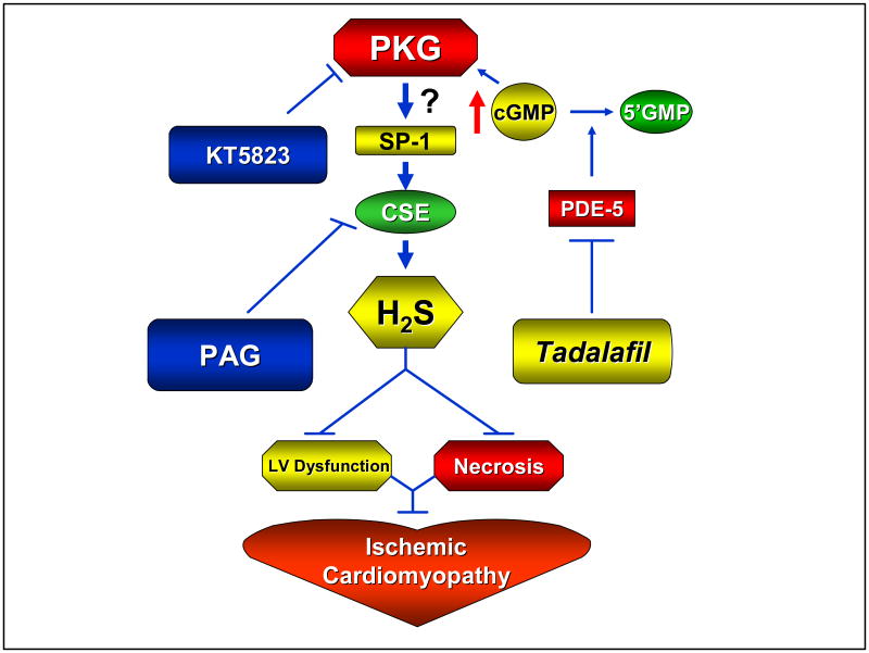
Background: Tadalafil is a novel long-acting inhibitor of phosphodiesterase-5. Because cGMP-dependent protein kinase (PKG) signaling plays a key role in cardioprotection, we hypothesized that PKG activation with tadalafil would limit myocardial ischemia/reperfusion (I/R) injury and dysfunction. Additionally, we contemplated that cardioprotection with tadalafil is mediated by hydrogen sulfide (H(2)S) signaling in a PKG-dependent fashion.
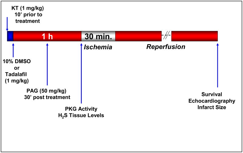
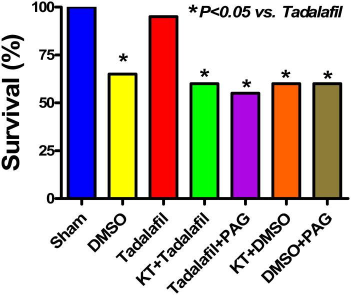
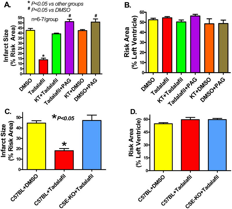
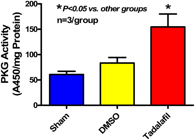
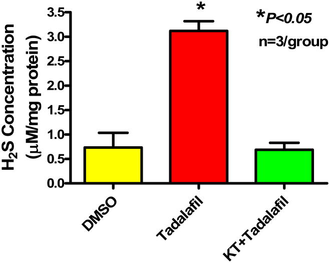
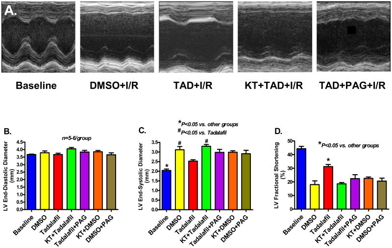
Methods and results: After baseline transthoracic echocardiography (TTE), adult ICR mice were injected i.p. with vehicle (10% DMSO) or tadalafil (1 mg/kg) with or without KT5823 (KT, PKG blocker, 1 mg/kg) or dl-propargylglycine (PAG, Cystathionine-gamma-lyase [CSE, H(2)S-producing enzyme] blocker; 50 mg/kg) 1 hour before coronary artery ligation for 30 minutes and reperfusion for 24 hours, whereas C57BL wild-type and CSE-knockout mice were treated with either vehicle or tadalafil. After reperfusion, TTE was performed and hearts were collected for infarct size (IS) measurement using TTC staining. Survival was increased with tadalafil (95%) compared with control (65%, P<0.05). Infarct size was reduced with tadalafil (13.2+/-1.7%) compared to vehicle (40.6+/-2.5%; P<0.05). KT and PAG abolished tadalafil-induced protection (IS: 39.2+/-1% and 51.2+/-2.4%, respectively) similar to genetic deletion of CSE (47.2+/-5.1%). Moreover, tadalafil preserved fractional shortening (FS: 31+/-1.5%) compared to control (FS: 22+/-4.8%, P<0.05). Baseline FS was 44+/-1.7%. KT and PAG abrogated the preservation of LV function with tadalafil by decline in FS to 17+/-1% and 23+/-3%, respectively. Compared to vehicle, myocardial H(2)S production was significantly increased with tadalafil and was abolished with KT.
Conclusions: PKG activation with tadalafil limits myocardial infarction and preserves LV function through H(2)S signaling.
Conflict of interest statement
Figures
References
-
- Kane LB, Klings ES. Present and future treatment strategies for pulmonary arterial hypertension: focus on phosphodiesterase-5 inhibitors. Treat Respir Med. 2006;5:271–82. - PubMed
-
- Ockaili R, Salloum F, Hawkins J, Kukreja RC. Sildenafil (Viagra) induces powerful cardioprotective effect via opening of mitochondrial KATP channels in rabbits. Am J Physiol Heart Circ Physiol. 2002;283:H1263–H1269. - PubMed
-
- Salloum FN, Ockaili RA, Wittkamp M, Marwaha VR, Kukreja RC. Vardenafil: a novel type 5 phosphodiesterase inhibitor reduces myocardial infarct size following ischemia/reperfusion injury via opening of mitochondrial KATP channels in rabbits. J Mol Cell Cardiol. 2006;40:405–11. - PubMed
-
- Salloum FN, Abbate A, Das A, Houser J, Mudrick CA, Qureshi IZ, Hoke NN, Roy SK, Brown WR, Prabhakar S, Kukreja RC. Sildenafil (Viagra) Attenuates Ischemic Cardiomyopathy and Improves Left Ventricular Function in Mice. Am J Physiol Heart Circ Physiol. 2008;294:H1398–406. - PubMed
-
- Padma-Nathan H. Efficacy and tolerability of tadalafil, a novel phosphodiesterase 5 inhibitor, in treatment of erectile dysfunction. Am J Cardiol. 2003;92:19M–25M. - PubMed
Publication types
MeSH terms
Substances
Grants and funding
LinkOut - more resources
Full Text Sources
