

Change in area of geographic atrophy in the Age-Related Eye Disease Study: AREDS report number 26
- PMID: 19752426
- PMCID: PMC6500457
- DOI: 10.1001/archophthalmol.2009.198
Change in area of geographic atrophy in the Age-Related Eye Disease Study: AREDS report number 26
Abstract
Objective: To characterize progression of geographic atrophy (GA) associated with age-related macular degeneration in AREDS as measured by digitized fundus photographs.
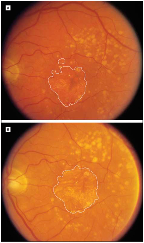
Methods: Fundus photographs from 181 of 4757 AREDS participants with a GA area of at least 0.5 disc areas at baseline or from participants who developed bilateral GA during follow-up were scanned, digitized, and evaluated longitudinally. Geographic atrophy area was determined using planimetry. Rates of progression from noncentral to central GA and of vision loss following development of central GA included the entire AREDS cohort.
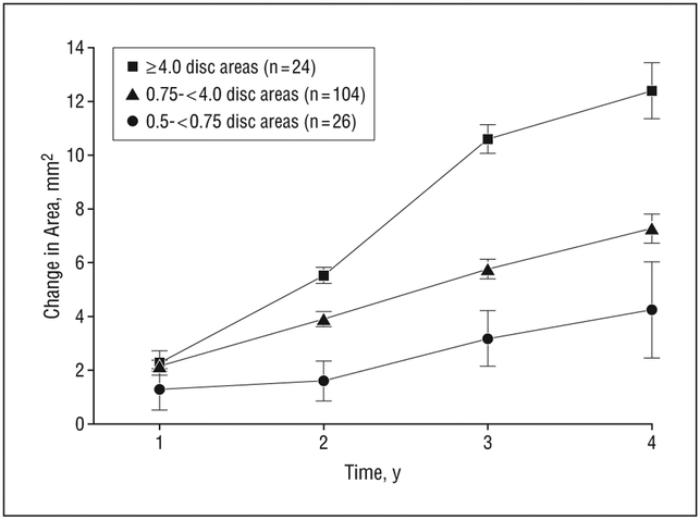
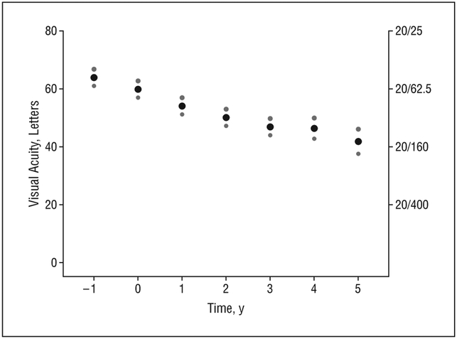
Results: Median initial lesion size was 4.3 mm(2). Average change in digital area of GA from baseline was 2.03 mm(2) (standard error of the mean, 0.24 mm(2)) at 1 year, 3.78 mm(2) (0.24 mm(2)) at 2 years, 5.93 mm(2) (0.34 mm(2)) at 3 years, and 1.78 mm(2) (0.086 mm(2)) per year overall. Median time to developing central GA after any GA diagnosis was 2.5 years (95% confidence interval, 2.0-3.0). Average visual acuity decreased by 3.7 letters at first documentation of central GA, and by 22 letters at year 5.
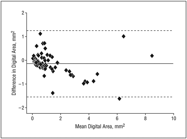
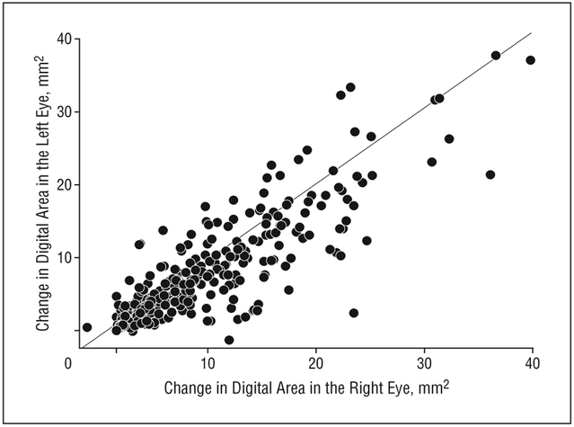
Conclusions: Growth of GA area can be reliably measured using standard fundus photographs that are digitized and subsequently graded at a reading center. Development of GA is associated with subsequent further growth of GA, development of central GA, and loss in central vision.
Figures
References
-
- Friedman DS, O’Colmain BJ, Muñoz B, et al.; Eye Diseases Prevalence Research Group. Prevalence of age-related macular degeneration in the United States. Arch Ophthalmol. 2004;122(4):564–572. - PubMed
-
- Lindblad AS, Clemons TE; Age-Related Eye Disease Study Research Group. Responsiveness of the National Eye Institute Visual Function Questionnaire to progression to advanced age-related macular degeneration, vision loss and lens opacity. AREDS Report No. 14. Arch Ophthalmol. 2005;123(9):1207–1214. - PMC - PubMed
-
- Sunness JS, Gonzalez-Baron J, Applegate CA, et al. Enlargement of atrophy and visual acuity loss in the geographic atrophy form of age-related macular degeneration. Ophthalmology. 1999;106(9):1768–1779. - PubMed
-
- Bellmann C, Jorzik J, Spital G, Unnebrink K, Pauleikhoff D, Holz FG. Symmetry of bilateral lesions in geographic atrophy in patients with age-related macular degeneration. Arch Ophthalmol. 2002;120(5):579–584. - PubMed
-
- Sunness JS, Applegate CA, Bressler NM, Hawkins BS Designing clinical trials for age-related geographic atrophy of the macula: enrollment data from the geographic atrophy natural history study. Retina. 2007;27(2):204–210. - PubMed
Publication types
MeSH terms
Substances
Grants and funding
LinkOut - more resources
Full Text Sources
Other Literature Sources
Medical
