. 2009 Sep;7(9):e1000196.
doi: 10.1371/journal.pbio.1000196. Epub 2009 Sep 15.

Loss of mitogen-activated protein kinase kinase kinase 4 (MAP3K4) reveals a requirement for MAPK signalling in mouse sex determination

Affiliations

Loss of mitogen-activated protein kinase kinase kinase 4 (MAP3K4) reveals a requirement for MAPK signalling in mouse sex determination

Debora Bogani et al. PLoS Biol. 2009 Sep.

Abstract

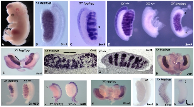
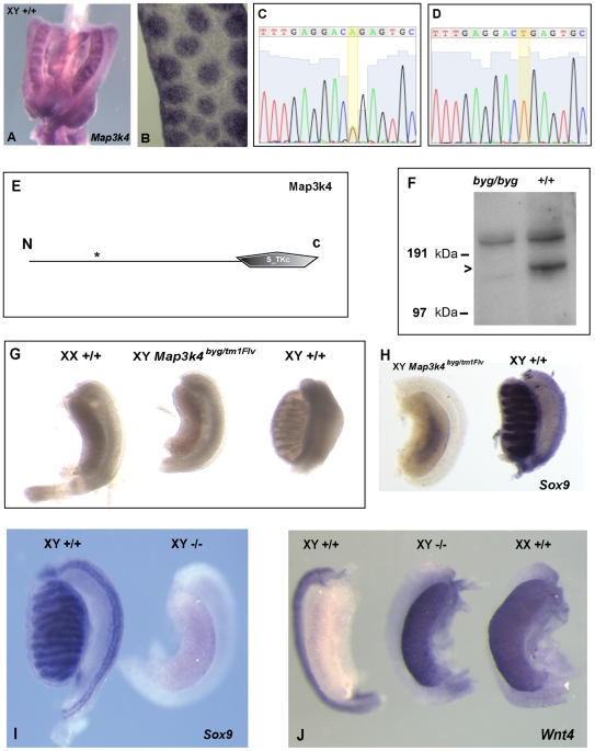
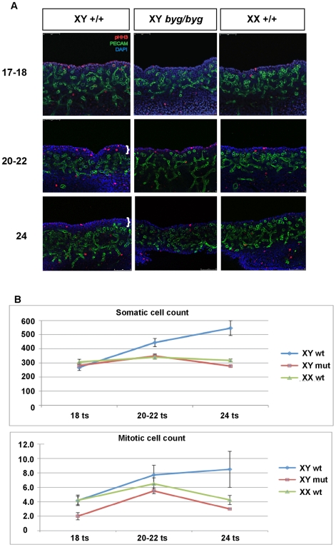
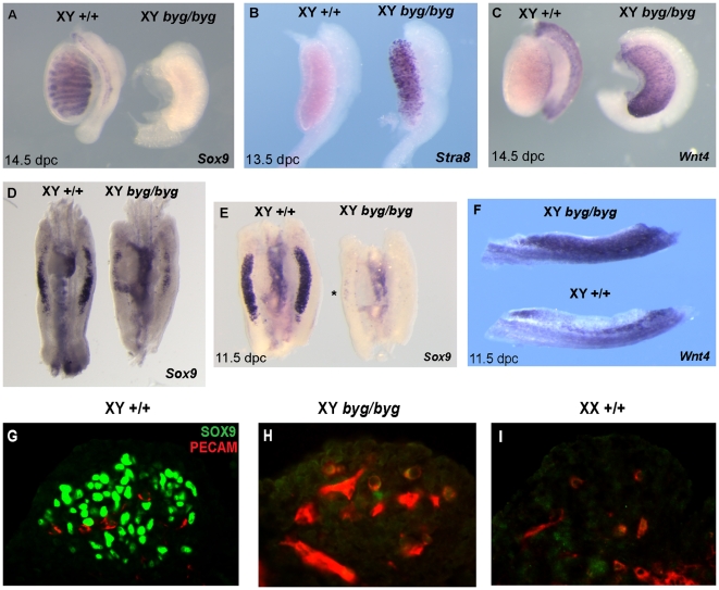
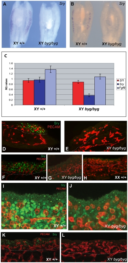
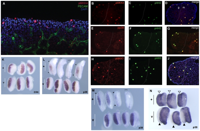
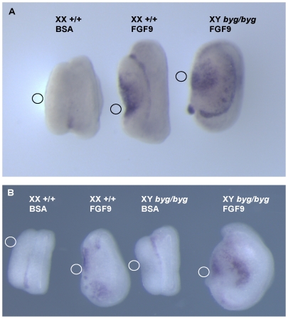
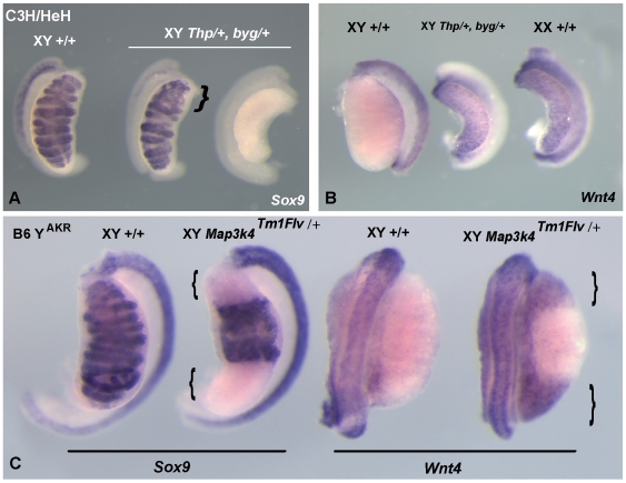
Sex determination in mammals is controlled by the presence or absence of the Y-linked gene SRY. In the developing male (XY) gonad, sex-determining region of the Y (SRY) protein acts to up-regulate expression of the related gene, SOX9, a transcriptional regulator that in turn initiates a downstream pathway of testis development, whilst also suppressing ovary development. Despite the requirement for a number of transcription factors and secreted signalling molecules in sex determination, intracellular signalling components functioning in this process have not been defined. Here we report a role for the phylogenetically ancient mitogen-activated protein kinase (MAPK) signalling pathway in mouse sex determination. Using a forward genetic screen, we identified the recessive boygirl (byg) mutation. On the C57BL/6J background, embryos homozygous for byg exhibit consistent XY gonadal sex reversal. The byg mutation is an A to T transversion causing a premature stop codon in the gene encoding MAP3K4 (also known as MEKK4), a mitogen-activated protein kinase kinase kinase. Analysis of XY byg/byg gonads at 11.5 d post coitum reveals a growth deficit and a failure to support mesonephric cell migration, both early cellular processes normally associated with testis development. Expression analysis of mutant XY gonads at the same stage also reveals a dramatic reduction in Sox9 and, crucially, Sry at the transcript and protein levels. Moreover, we describe experiments showing the presence of activated MKK4, a direct target of MAP3K4, and activated p38 in the coelomic region of the XY gonad at 11.5 d post coitum, establishing a link between MAPK signalling in proliferating gonadal somatic cells and regulation of Sry expression. Finally, we provide evidence that haploinsufficiency for Map3k4 accounts for T-associated sex reversal (Tas). These data demonstrate that MAP3K4-dependent signalling events are required for normal expression of Sry during testis development, and create a novel entry point into the molecular and cellular mechanisms underlying sex determination in mice and disorders of sexual development in humans.


Conflict of interest statement

The authors have declared that no competing interests exist.

Figures

Figure 1
Figure 1. Identification of the byg mutant.
(A) Line RECB/31 (boygirl, byg) mutant embryo dissected during the screen at 13.5 dpc. The embryo shown here has exencephaly, spina bifida, and mild oedema. On the mixed genetic background used for the forward genetic screen, the boygirl XY gonadal phenotypes observed at 13.5/14.5 dpc ranged from testis-shaped gonads that lacked cords (B), to testes with abnormal cord morphology and “missing” cords (arrowhead, C) through to XY gonads with an overt ovarian morphology (right, D). All gonads shown here were hybridised with a Sox9 probe to assess cord structure and differentiation of Sertoli cells. Note the higher than normal (with respect to ovary development) levels of Sox9 transcript in the boygirl XY “ovary” in (D) and the absence of Sox9 from the poles of this gonad (arrowheads, D), a characteristic of ovotestis development. Wild-type littermate gonads from unaffected XY and XX embryos are shown for comparison. (E–G) After further out-crossing of the byg mutation to C3H/HeH for mapping purposes, XY byg/byg gonads exhibited further signs of ovotestis formation. For example, expression of the germ cell marker Oct4 at 13.5 dpc revealed a cord-like pattern only in the centre of the gonad shown here (bracket, E), made clearer by sectioning (bracket, F). Note also the thickened capsule over the central testicular area. Control gonads exhibited robust cord formation throughout their length (G). (H) XX byg/byg gonads exhibited a normal pattern of Oct4 expression. (I) The poles of XY byg/byg gonads were also characterised by loss of male-specific genes, such as the Leydig cell marker 3β-HSD (bracket). (J) Abnormal expression of female-specific marker Wnt4 at the gonadal poles (arrowheads next to gonad on left) at 14.5 dpc. (K) Normal expression of Wnt4 in XX byg/byg gonad. (L) Absence of expression of meiotic marker Stra8 in wild-type XY gonad. (M) Prominent Stra8 expression in XX byg/byg gonad. (N) Expression of Stra8 at gonadal poles in XY byg/byg gonad (brackets).
Figure 2
Figure 2. The byg phenotype is caused by a point mutation in Map3k4.
(A) WMISH of 12.5 dpc XY gonads with a Map3k4 probe revealing widespread expression, including in newly formed testis cords. (B) Longitudinal section through male gonad at 13.5 dpc, after WMISH, showing Map3k4 expression in testis cords. (C, D) Sequence traces from heterozygous (byg/+, C) and homozygous (byg/byg, D) individuals reveal an A to T transversion at nucleotide position 1,144 of the Map3k4 open reading frame of the byg allele. (E) This mutation replaces an arginine with a premature stop codon (asterisk) at amino acid position 382 of the 1,597 amino acid MAP3K4 protein. The predicted truncated protein lacks the critical kinase domain (S_TKc) and, therefore, any MAPKKK function. (F) Western blotting of protein extracted from byg/byg and +/+ embryos shows absence of full-length (180 kDa) MAP3K4 in mutant homozygotes. The position of size markers is shown on the left. The upper band found in both lanes is due to cross-reaction of the antibody with an unrelated protein. (G) A genetic complementation test was performed to confirm that homozygosity for the Map3k4 point mutation caused the byg gonadal phenotype. XY mutant embryos heterozygous for both the Map3k4byg and targeted Map3k4tm1Flv alleles were dissected at 14.5 dpc and contained gonads with an overt ovarian morphology (central gonad). XX and XY littermate controls are also shown. (H) Hybridisation of a Sox9 probe to a doubly heterozygous XY gonad (left) reveals little expression of the Sertoli cell marker, in contrast to an XY control (right). (I) XY gonad from 14.5 dpc embryo homozygous for the Map3k4tm1Flv allele (−/−) exhibits overt ovarian morphology and an absence of Sox9 (right), in contrast to XY littermate control (left). (J) XY gonad from homozygous knockout embryo (−/−) also expresses high levels of Wnt4 (central gonad). XY and XX control gonads are shown on left and right, respectively.
Figure 3
Figure 3. Reduced gonadal growth in XY byg/byg embryos between 11.5 and 12.25 dpc.
(A) Somatic cell proliferation in the coelomic region of control and XY byg/byg gonads was analysed by confocal imaging of wholemount organs after immunostaining with anti-PECAM (green) and anti-phospho-histone H3 (red) antibodies. Cell nuclei were visualised using DAPI staining (blue). All gonads were staged accurately by counting ts (ts number shown on left). The coelomic growth zone characteristic of XY gonads is shown with white brackets in the 22 ts and 24 ts samples. This thickened zone of proliferating somatic cells is not found in XY byg/byg or XX +/+ gonads at any of the stages analysed. (B) Counts in the coelomic region of total number of somatic cells and mitotic (pHH3-positive) cells in XX/XY wild-type (wt) and XY byg/byg (mut) gonads at the stages shown in (A). Cell counts were performed on limited numbers of XX byg/byg gonads at 18 ts and 20–22 ts. Numbers were comparable with wild-type XX and XY byg/byg samples (unpublished data). For details of cell counting methodology and statistical tests see Materials and Methods and Table 1.
Figure 4
Figure 4. Analysis of Sox9, Wnt4, and Stra8 expression reveals gonadal sex reversal in XY byg/byg embryos.
(A) After back-crossing of the byg mutation to C57BL/6J for at least two generations, XY byg/byg gonads expressed negligible levels of Sox9 (left) in comparison to control XY gonads (right) at 14.5 dpc. (B) At 13.5 dpc, XY byg/byg gonads express Stra8, a marker of meiotic germs cells usually restricted to the ovary. (C) Wnt4, a marker of ovarian somatic cells, is expressed in mutant XY gonads (right), in contrast to control gonads (left). (D) On the C3H/HeH background Sox9 transcription is detected at lower levels in the XY byg/byg gonad at 11.5 dpc (right) when compared to wild-type controls (left). Signal in the mutant gonad appears concentrated towards the centre. (E) After backcrossing to C57BL/6J, Sox9 is expressed abundantly in the XY wild-type gonad at 11.5 dpc (left) but is now absent from XY byg/byg gonads, apart from just a few cells visible in the central region (asterisk, right). (F) XY byg/byg gonads at 11.5 dpc express the ovarian somatic cell marker Wnt4 (top), in contrast to an XY control gonad (bottom). (G–I) Immunostaining of a transverse section from a control C57BL/6J XY gonad at 11.5 dpc with anti-PECAM (germ cell, red) and anti-SOX9 (pre-Sertoli cell, green) antibodies reveals large numbers of somatic cell nuclei (G), in contrast to XY byg/byg (H), and XX wild-type control (I), in which SOX9 protein is not detected at significant levels.
Figure 5
Figure 5. Loss of Sry transcript and protein expression in XY byg/byg gonads at 11.5 dpc on C57BL/6J.
(A) At the 17 ts stage, Sry transcript is detected throughout an XY control gonad (left) but is absent from the C57BL/6J XY byg/byg gonad (right). (B) By 19 ts, the signal is diminishing in the XY control gonad (left) and is still not detectable in the XY byg/byg gonad (right). (C) qRT-PCR analysis of Sry, Sf-1, and Fgf9 transcription in XY +/+ and byg/byg gonads at 11.5 dpc (17–18 ts). Error bars for the relative quantitation (RQ) values represent variation across four biological replicates for each genotype and three technical replicates for each sample. The 2.8-fold reduction in Sry levels in XY byg/byg gonads is significant (p = 0.02) based on a t-test calculated using average dCT values for the above genes and those of Hprt1. Differences in the levels of Sf-1 (p = 0.28) and Fgf9 (p = 0.23) were not significant, although there is a trend for reduced expression of Fgf9 in byg/byg. (D) Immunostaining of a longitudinal section of control XY gonad at 17 ts with anti-SRY (green) and anti-PECAM (red) antibodies reveals abundant expression of SRY in somatic cell nuclei of the gonad. Tissue beneath the dotted white line in this and subsequent images is mesonephric. (E) In contrast, very few SRY-positive cells are detectable in a longitudinal section from a XY byg/byg gonad at the same stage. (F) Confocal imaging of a control XY gonad after wholemount immunostaining with anti-SRY and anti-PECAM antibodies reveals large numbers of SRY-positive somatic cells, in contrast to XY byg/byg (G) and control XX (H) gonads. (I) High magnification confocal image of wild-type gonad at 11.5 dpc showing large numbers of SRY-positive somatic cells (green) and germ cells (red). (J) Confocal image of SRY expression in XY byg/byg gonad at 11.5 dpc generated using the same settings as in (I). Note the greatly reduced number of SRY-positive cells and the reduction in signal intensity in the mutant gonad. (K) A wild-type XY gonad at 11.0 dpc (13 ts) showing SRY-positive cells (green) amongst germ cells (red). (L) In contrast, no SRY-positive cells are detected at 11.0 dpc in an XY byg/byg gonad.
Figure 6
Figure 6. MAPK signalling and XY gonad development.
(A–G) Gonadal expression of three activated MAPK signalling components was examined: phospho-MKK4 (pMKK4), a product of MAP3K4-mediated phosphorylation of MKK4, phospho-p38 (pp38), and phospho-MKK7 (pMKK7). (A) Anti-pMKK4 antibody detects activated MKK4 (red) in a number of somatic cells in the developing XY gonad at 11.5 dpc (21 ts) after wholemount immunostaining. Note the concentration of pMKK4-positive somatic cells at the gonadal periphery in the coelomic region. Germ cells are marked by anti-PECAM antibody (green) and cell nuclei by DAPI. (B–D) Transverse sections of 21 ts XY gonad showing co-expression of pMKK4 and the mitotic marker pHH3 in cells at the gonadal periphery. The gonad (left) is separated from the mesonephros (right) by a dotted white line. (E–G) Transverse sections of 21 ts XY gonad revealing co-expression of activated p38 (pp38) and pHH3 in cells at the gonadal periphery. (H–J) Transverse sections showing similar co-expression of pMKK7 and pHH3. (K–N) The effect of two specific inhibitors of MAPK signalling on XY gonad development in vitro was studied. (K) Culture of wild-type XY gonads from 11.5 dpc for 48 h in the presence anti-ERK inhibitor U0126 (+, upper three gonads in panel) has no obvious effect on Sox9 expression in comparison to gonads cultured with vehicle control (v, lower three gonads). Testis cord formation in treated samples is, however, not as pronounced as in controls. (L, M) Culture of wild-type XY gonads in the presence of p38 inhibitor SB202190 (+, upper rows of gonads) results in striking, but variable, alterations to Sox9 expression patterns in contrast to vehicle control cultures (v, lower rows), ranging from loss of transcription in the gonadal poles (asterisks) to complete absence of transcription (gonads beneath brackets). (N) Analysis of XY gonads cultured in SB202190 also reveals up-regulation of Wnt4 transcription (open arrowheads, upper row) in contrast to vehicle controls, which lack Wnt4 (black arrowheads, lower row). These data suggest that, at least partial, XY gonadal sex reversal is caused by inhibition of p38 from 11.5 dpc. There were no signs of tissue necrosis or excessive cell death during these experiments. All gonads were from embryos on the C57BL/6J background.
Figure 7
Figure 7. Exogenous FGF9 can activate Sox9 transcription in gonads lacking functional MAP3K4.
(A) Culture of a wild-type XX gonad (left) from 11.5 dpc for 48 h in the presence of a bead coated in BSA does not activate Sox9 transcription based on in situ hybridisation analysis. In contrast, a bead coated in FGF9 does result in up-regulation of Sox9 in both XX control gonads (centre) and XY byg/byg gonads (right). The small circle adjacent to each gonad shows the approximate position of the bead during culture. (B) In a separate experiment, FGF9-coated beads again activate Sox9 transcription in wild-type XX and XY byg/byg gonads, but a bead coated in BSA does not activate Sox9 transcription in XY byg/byg gonads. Note the increased size of gonads, both XX and XY, after exposure to exogenous FGF9. All gonads were from embryos on the C57BL/6J background.
Figure 8
Figure 8. Hemizygosity for Map3k4 contributes to Tas.
(A, B) A genetic complementation test demonstrates that Map3k4 resides in the Thp deletion of proximal mouse Chromosome 17. XY embryos doubly heterozygous for the Map3k4byg allele and the Thp deletion (XY Thp/+, byg/+), on the C3H/HeH background, exhibit a range of defects of gonad development similar to byg/byg homozygotes, including testes with regions lacking clear cord morphology (bracket, A) and XY gonads with an overt ovarian appearance that lack Sox9 transcription (right-hand side gonad, A). Sex-reversed XY gonads express Wnt4, a marker of ovary development (central gonad, B), in contrast to XY controls. (C) Ovotestis development in embryos heterozygous for the Map3k4tm1Flv knockout allele, on the C57BL/6J XYAKR background (B6YAKR), characterised by gonadal poles (brackets) lacking markers of testis development (Sox9), and expressing markers of ovary development (Wnt4).

References

    1. Brennan J, Capel B. One tissue, two fates: molecular genetic events that underlie testis versus ovary development. Nat Rev Genet. 2004;5:509–521. - PubMed
    1. Gubbay J, Collignon J, Koopman P, Capel B, Economou A, et al. A gene mapping to the sex-determining region of the mouse Y chromosome is a member of a novel family of embryonically expressed genes. Nature. 1990;346:245–250. - PubMed
    1. Koopman P, Münsterberg A, Capel B, Vivian N, Lovell-Badge R. Expression of a candidate sex-determining gene during mouse testis differentiation. Nature. 1990;348:450–452. - PubMed
    1. Koopman P, Gubbay J, Vivian N, Goodfellow P, Lovell-Badge R. Male development of chromosomally female mice transgenic for Sry. Nature. 1991;351:117–121. - PubMed
    1. Lovell-Badge R, Robertson E. XY female mice resulting from a heritable mutation in the murine primary testis determining gene, Tdy. Development. 1990;109:635–646. - PubMed

Publication types

Substances