

doi: 10.4103/0019-5413.45333.
Benign self-limiting cystic lesion after lower end radius fracture in a child
Affiliations
- PMID: 19753191
- PMCID: PMC2739504
- DOI: 10.4103/0019-5413.45333
Item in Clipboard
Benign self-limiting cystic lesion after lower end radius fracture in a child
Indian J Orthop.
2009 Jan.
Abstract
Post-traumatic cystic lesion is usually found adjacent to a healing torus fracture. It is typically asymptomatic and appears just proximal to the fracture line within the area of subperiosteal new bone formation. We report one such cyst in a 7 year old boy, with a brief review of literature to highlight the occurrence of such benign self limiting cystic lesions of lower end radius fracture.
Keywords: Benign cyst; distal radius; torus fracture.
Conflict of interest statement
Figures

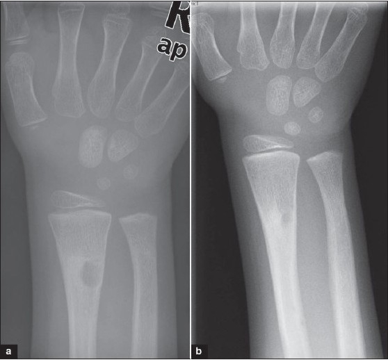
Ateroposterior(a) and lateral(b) X-rays of the forearm with wrist shows Greenstick fracture of the distal radius, with dorsal angulation. Anteroposterior (c) and lateral (d) xray at 3 weeks follow up shows faintly defined cystic area within the subperiosteal callus, just proximal to the fracture site.
Anteroposterior X-ray (a) of forearm with wrist at 3 month follow-up shows a well defined cystic lesion at the original site of the lesion within the newly formed subperiosteal bone. (b) X-ray at six month follow-up shows complete resolution of the cyst.

T1WI of MRI (a) at three months follow-up shows area of increased signal with a density similar to that of fat adjacent to the fracture and corresponding to the area of the cystic lesion. Two other coincidental areas of increased signal of unknown significance seen in proximal radial shaft. Fat suppressed image (b) confirming fatty composition of the cyst.
References
-
- Davids JR, Graner KA, Mubarak SJ. Post-fracture lipid inclusion cyst: A case report. J Bone Joint Surg Am. 1993;75:1528–32. - PubMed
-
- Garcia-Alvarez F, Bello M, Albareda J, Seral F. Transient cyst-like cortical defect following radius fracture in children. Int Pediatr. 1999;14:179.
-
- Dürr HR, Lienemann A, Stäbler A, Küehne JH, Refior HJ. MRI of post-traumatic cyst-like lesions of bone after a greenstick fracture. J Eur Radiat. 1997;7:1218–20. - PubMed
-
- Pfister-Goedecke L, Braune M. Cyst-like cortical defects following fractures in children. Pediatr Radiol. 1981;11:83–6. - PubMed
LinkOut - more resources
Full Text Sources