. 2009 Sep 15;4(9):e7028.
doi: 10.1371/journal.pone.0007028.

Decorin core protein (decoron) shape complements collagen fibril surface structure and mediates its binding

Affiliations

Decorin core protein (decoron) shape complements collagen fibril surface structure and mediates its binding

Joseph P R O Orgel et al. PLoS One. .

Abstract

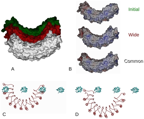
Decorin is the archetypal small leucine rich repeat proteoglycan of the vertebrate extracellular matrix (ECM). With its glycosaminoglycuronan chain, it is responsible for stabilizing inter-fibrillar organization. Type I collagen is the predominant member of the fibrillar collagen family, fulfilling both organizational and structural roles in animal ECMs. In this study, interactions between decoron (the decorin core protein) and binding sites in the d and e(1) bands of the type I collagen fibril were investigated through molecular modeling of their respective X-ray diffraction structures. Previously, it was proposed that a model-based, highly curved concave decoron interacts with a single collagen molecule, which would form extensive van der Waals contacts and give rise to strong non-specific binding. However, the large well-ordered aggregate that is the collagen fibril places significant restraints on modes of ligand binding and necessitates multi-collagen molecular contacts. We present here a relatively high-resolution model of the decoron-fibril collagen complex. We find that the respective crystal structures complement each other well, although it is the monomeric form of decoron that shows the most appropriate shape complementarity with the fibril surface and favorable calculated energies of interaction. One molecule of decoron interacts with four to six collagen molecules, and the binding specificity relies on a large number of hydrogen bonds and electrostatic interactions, primarily with the collagen motifs KXGDRGE and AKGDRGE (d and e(1) bands). This work helps us to understand collagen-decorin interactions and the molecular architecture of the fibrillar ECM in health and disease.


Conflict of interest statement

Competing Interests: Joseph Orgel is a member of the academic_editorial Board of PLoS ONE and Section academic_editor for Biochemistry.

Figures

Figure 1
Figure 1. Consensus collagen sequences for decorin binding sites and decoron binding conformational orientations.
A) Rabbit ventral skin, stained with Cupromeronic blue to demonstrate the proteoglycan filaments (which are about one D period apart) orthogonal to the collagen fibrils and subsequently with uranyl acetate to delineate the a–e banding pattern. B) The consensus decoron binding collagen sequences are shown here as coloured bands on a representation of the type I collagen microfibril , collagen N→C direction runs from bottom to top: Yellow = D∼2.65 (AOGDKGEAGPSG) e 2-band site (partially accessible) Cyan = D∼2.74 (AOGDRGEOGPOG) e 1-band site (partially accessible) Blue = D∼3.74 (AKGDRGETGPAG) e 1-band site (fully accessible) Red = D∼3.87 (KNGDRGEOGPAG) d-band site (fully accessible). The positions of two non-binding sites used as negative controls in the molecular docking calculations are indicated as nbs1 and nbs2. The central representation is viewed from the exterior of the fibril surface, the left and right views are from within the fibril (red arrows point to fibril exterior). C) Electrostatic rendering of accessible surface area of a decoron in the Dec N→C or Dec C→N dimer (left) or monomer (right) conformations. For the Dec N→C conformation, the N-terminus is leftmost for the monomer, and in the central section of the dimer (inside of the dimer interface).
Figure 2
Figure 2. Molecular packing at the collagen fibril surface and decoron-fibril binding.
A) Schematic, composite representation of a decoron molecule bound to the fibril surface (e 1-band site) in the Dec N→C orientation; collagen monomers 1–4 from each microfibril are labelled. The four monomers in closest association with the docked decoron monomer are surface rendered in red (the decoron molecule is surface rendered in blue). B) Molecular packing structure of collagen monomers at the fibril surface at ∼0.74 D (e 1-band site). 1: Represents the “common” arrangement of monomers around most of the fibril surface. 2: The maximum “wide” model.
Figure 3
Figure 3. Decoron docking at the fibril surface for the common and wide arrangements at the d and e1 binding sites.
Side chains of the amino acid residues involved in the decoron/collagen interface are shown in red (collagen) or blue (decoron). Candidate interacting residues must be capable of forming at least one hydrogen bond, and to be less than 4 Å from a residue in the other molecule. Possible hydrogen bond interactions are shown in panels A–D for different model arrangements, all are in the Dec N→C orientation (see also Table S1). A) e 1-band site, common conformation, decoron monomer. B) e 1-band site, wide conformation, decoron monomer. C) d-band site, common conformation, decoron dimer. D) d-band site, wide conformation, decoron dimer. E) Decoron molecule docked at the common fibril surface model in the e 1-band (blue). Oligosaccharide binding residues are shown, as is the AGAG chain binding N-terminal sequence (yellow). Note that the decoron molecule is tilted (∼8.5 degrees relative to the lateral plane of the collagen fibril) in its final energy minimized conformation, and the carbohydrate binding amino acid residues all appear to be fully accessible. In all A–E panels every other LRR is coloured green for reference (the first being LRR-1 at the N-term, then LRR-2 is gray, LRR-3 is green etc; the region N-terminal to LRR-1, including part of the capping structure, is shown in gray).
Figure 4
Figure 4. Curvature of the decoron monomer.
A) Decoron's radius of curvature is only modestly changed upon binding fibril surface. The initial (crystal) structure and the wide and common conformation bound models of decoron were superimposed and then stacked along the molecules height axis to compare their overall shape and curvature B) Electrostatic rendering of decoron for each model referred to above. Note that for the common and wide bound models of decoron have more even, bracket shaped interior but are still not the highly arched shapes previously envisioned. C) In contrast to A and B, the decoron model is based on the ribonuclease inhibitor structure rather than the decoron crystal structure is highly curved. Here it is shown attempting to dock with the fibril surface as for Figures 2–3. Note the substantial molecular overlap that occurs when the decoron is docked to an individual collagen molecule, with the neighboring collagen molecules at the fibril surface. D) As C, except: the ribonuclease inhibitor based decoron molecule has been placed to avoid steric clashes, note that the receptor-ligand interface appears substantially less engaged than that seen in Figures 2–3.
Figure 5
Figure 5. Homologous residues that contribute most to the energy of association.
A) Homology between bovine decoron and bigylcan rendered as dark green for identical residues and light green for similar residues. Residues that are not common between the two proteins are rendered gray. B) Map of the decoron residues that most contribute to the energy of association with collagen for the 4 primary collagen receptors studied here (common and wide conformations for the d and e1 band sites), see Table S1 for reference. The darker shade of yellow corresponds to residues that are ranked as contributing highly to the ligand-receptor interaction (methods, Table S1). C) As for A), except comparison is between rat and bovine decoron sequences.
Figure 6
Figure 6. Free energy of collagen-ligand association for fibril surface receptor models.

References

    1. Scott JE, Stockwell RA. Cartilage elasticity resides in shape module decoran and aggrecan sumps of damping fluid: implications in osteoarthrosis. J Physiol (Lond.) 2006;574:643–50. - PMC - PubMed
    1. Daley W, Peters S, Larsen M. Extracellular matrix dynamics in development and regenerative medicine. Journal of cell science. 2008;121:255–64. - PubMed
    1. Sweeney S, et al. Candidate cell and matrix interaction domains on the collagen fibril, the predominant protein of vertebrates. Journal of biological chemistry. 2008;283:21187–21197. - PMC - PubMed
    1. Perumal S, Antipova O, Orgel JPRO. Collagen fibril architecture, domain organization, and triple-helical conformation govern its proteolysis. Proc Natl Acad Sci U S A. 2008;105:2824–2829. - PMC - PubMed
    1. Scott JE. Supramolecular organization of extracellular matrix glycosaminoglycans, in vitro and in the tissues. The FASEB journal. 1992;6:2639–45. - PubMed

Publication types