Comparative analysis of gene transfer to human and rat retinal pigment epithelium cell line by a combinatorial use of recombinant adeno- associated virus and ultrasound or/and microbubbles
- PMID: 19754469
- PMCID: PMC5632498
- DOI: 10.17305/bjbms.2009.2802
Comparative analysis of gene transfer to human and rat retinal pigment epithelium cell line by a combinatorial use of recombinant adeno- associated virus and ultrasound or/and microbubbles
Abstract
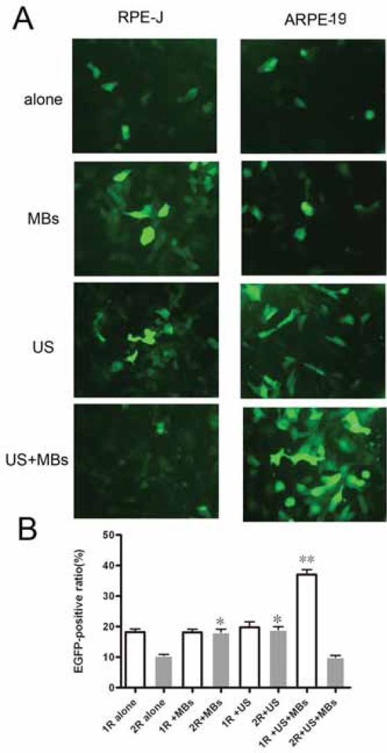
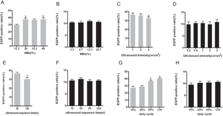
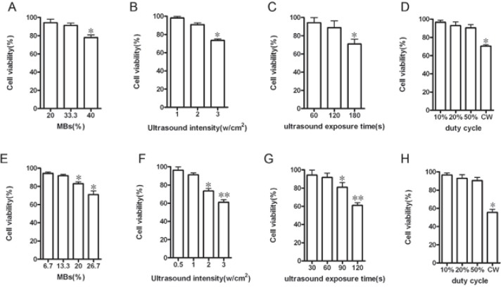
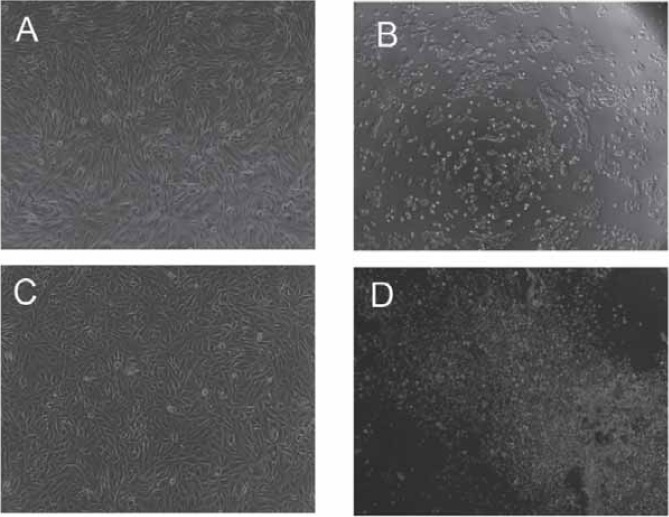
Ultrasound-targeted microbubble destruction has been utilized to deliver a drug/gene into cells in both in vitro and in vivo studies. This work was performed to investigate the feasibility of gene transfer to human retinal pigment epithelium cell line(ARPE-19) and rat retinal pigment epithelium cell line(RPE-J) by a combinatorial use of recombinant adeno-associated virus (rAAV) and ultrasound (US) or/and microbubbles (MBs) and compare the difference between them. Different doses of serotype 2 rAAV encoding a enhanced green fluorescent protein (rAAV2-EGFP) gene and MBs was administered to ARPE-19 and RPE-J cells under different US conditions. Transfection efficiency and cell viability were assessed by fluorescence microscopy, flow cytometry (FCM) analysis, trypan blue staining. The results indicated that US and MBs could respectively improve rAAV2-mediated gene transfer to RPE-J cells, but neither US nor MBs could do so in ARPE-19 cells. US plus MBs could significantly enhance rAAV2-mediated gene transfer to ARPE-19 cells, however, the same effects were not seen in RPE-J cells. These findings demonstrated it is not always coincident that US, MBs and US plus MBs exert the similar effects on gene transfer in vitro RPE cells. So, it is necessary to choose appropriate RPE cell line for the study of US or/and MBs-mediated rAAV gene transfer in retinal gene therapy.
Figures
References
-
- West K.A, Yan L, Miyagi M, et al. Proteome survey of proliferat-ing and differentiating rat RPE-J cells. Exp.Eye.Res. 2001;73:479–491. - PubMed
-
- Pang J.J, Lauramore A, Deng W.T, et al. Comparative analysis of in vivo and in vitro AAV vector transduction in the neonatal mouse retina: Effects of serotype and site of administration. Vision Res. 2008;48:377–385. - PubMed
-
- Bekeredjian R, Grayburn P.A, Shohet R.V. Use of ultrasound contrast agents for gene or drug delivery in cardiovascular medicine. J.Am.Coll. Cardiol. 2005;45:329–335. - PubMed
-
- Liu Y, Miyoshi H, Nakamura M. Encapsulated ultrasound mi-crobubbles: therapeutic application in drug/gene delivery. J. Control Release. 2006;114:89–99. - PubMed