Review
. 2009 Sep;231(1):59-87.
doi: 10.1111/j.1600-065X.2009.00816.x.

The functional network of ion channels in T lymphocytes

Affiliations
Review

The functional network of ion channels in T lymphocytes

Michael D Cahalan et al. Immunol Rev. 2009 Sep.

Abstract

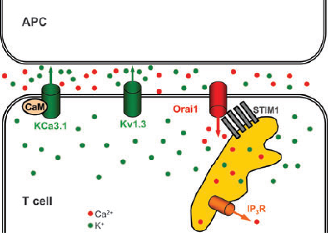
For more than 25 years, it has been widely appreciated that Ca2+ influx is essential to trigger T-lymphocyte activation. Patch clamp analysis, molecular identification, and functional studies using blockers and genetic manipulation have shown that a unique contingent of ion channels orchestrates the initiation, intensity, and duration of the Ca2+ signal. Five distinct types of ion channels--Kv1.3, KCa3.1, Orai1+ stromal interacting molecule 1 (STIM1) [Ca2+-release activating Ca2+ (CRAC) channel], TRPM7, and Cl(swell)--comprise a network that performs functions vital for ongoing cellular homeostasis and for T-cell activation, offering potential targets for immunomodulation. Most recently, the roles of STIM1 and Orai1 have been revealed in triggering and forming the CRAC channel following T-cell receptor engagement. Kv1.3, KCa3.1, STIM1, and Orai1 have been found to cluster at the immunological synapse following contact with an antigen-presenting cell; we discuss how channels at the synapse might function to modulate local signaling. Immuno-imaging approaches are beginning to shed light on ion channel function in vivo. Importantly, the expression pattern of Ca2+ and K+ channels and hence the functional network can adapt depending upon the state of differentiation and activation, and this allows for different stages of an immune response to be targeted specifically.


Figures

Fig. 1
Fig. 1. Five types of ion channels in T lymphocytes
From top to bottom: whole-cell current fingerprints (10, 50, 64, 111, 126); molecular identities of membrane-spanning subunits and accessory subunits or domains; and examples of channel blockers including ShK sea anemone peptide toxin (lower left) showing the critical lysine 22 in red (38). From left to right: voltage-gated K+ channel (Kv1.3), with structure of Kvβ2 (227); Ca2+-acti-vated K+ channel (KCa3.1), with CaM structure (PDB 1up5); CRAC channel (Orai1 + STIM1), EF-SAM domain structure of STIM1 (292); MIC channel (TRPM7), kinase domain structure (293); and Clswell of uncertain molecular composition. CRAC, Ca2+-release activating Ca2+ channel; STIM, stromal interacting molecule; MIC, Mg2+-inhibited Ca2+-permeable current; Clswell, swelling-activated Cl channel; CaM, calmodulin.
Fig. 2
Fig. 2. Biophysical characteristics of K+ channels in T lymphocytes
(A) Voltage-gated K+ current (from 22, 147). From left to right: whole-cell currents in response to step depolarization to varying potentials, voltage dependence of channel opening (red box shows normal range of resting membrane potential near the foot of the channel activation curve); outside-out patch single-channel current in response to a voltage ramp stimulus. (B) Ca2+-activated K+ current (from 50). From left to right: whole cell currents increasing as Ca2+ enters the cytosol; Ca2+ dependence of channel opening (red box shows normal range of cytosolic Ca2+ levels at rest, indicating lack of KCa3.1 channel activation until after the Ca2+ signal is initiated); single channels in inside-out patch exposed to varying Ca2+ concentrations.
Fig. 3
Fig. 3. CRAC channel
(A) Native CRAC current (yellow highlighted trace) in Jurkat T cells activated during passive store depletion (from 64). (B) Amplified CRAC current in S2 cells co-transfected with Stim + Orai (from 78). Note the difference in current scales and the increase in current size when the extracellular Ca2+ concentration is increased from 2 to 20 mM. Amplified CRAC currents represent approximately 105 channels per cell or 100 functional channels per µm2 of membrane surface in the overexpression system. (C) Orai sequence from first to second transmembrane segments. Residues conserved in Orai, Orai1, Orai2, and Orai3 are shown in bold. E180 Orai corresponds to E106 in Orai1. (D) Altered ion selectivity resulting from a conservative point mutation of Orai from glutamate to aspartate at position 180. Point mutation of this critical glutamate converts CRAC current from inwardly rectifying and Ca2+ selective to outwardly rectifying and monovalent cation selective (yellow highlighted trace). Stim was co-expressed to amplify CRAC currents (recordings from 87). CRAC, Ca2+-release activating Ca2+ channel; Stim, stromal interacting molecule.
Fig. 4
Fig. 4. Ion channels and Ca2+ signaling in T cells
Signaling pathway from TCR engagement to gene expression in nucleus. Major ion channel types are color coordinated according to ion selectivity, Orai1 (red), Kv1.3 and KCa3.1 (green), cation non-selective TRPM7 and IP3R channels (orange), and Clswell (violet). Dots and arrows correspond to ions and fluxes with the same color code. STIM1 is shown (gray) with a Ca2+ ion bound to its EF-hand within the ER lumen under basal conditions with the ER Ca2+ store filled. Ion channels and functions include (clockwise from right): Kv1.3 maintains the resting membrane potential and participates in RVD; Clswell triggers RVD by opening in response to cell swelling; KCa3.1 hyperpolarizes the membrane potential when cytosolic Ca2+ rises; Orai1 embodies the pore-forming subunit of the CRAC channel and is activated by STIM1 following ER Ca2+ store depletion (dotted line); TRPM7, activated by PIP2 and inhibited by Mg2+ inside, may regulate Mg2+ homeostasis in the cell. Proximal signaling events inside the cell following presentation of antigen include the following (counterclockwise from top right): TCR engagement of peptide–MHC, activation of tyrosine kinases (TK: Lck, Fyn, and ZAP-70), and phospholipase-C-γ (PLCγ), resulting in the cleavage of PIP2 to generate the second messengers IP3 and DAG; IP3-induced Ca2+ release; depletion-induced mobilization of STIM1 and activation of Ca2+ influx through Orai1 subunits. Post-Ca2+ events include activation of KCa3.1 via prebound CaM, activation of calcineurin via CaM, accumulation of dephosphorylated NFAT subunits in the nucleus, binding to DNA promoter regions, and altered gene expression. Functionally significant changes in ion channel expression and Ca2+ signaling are indicated by arrows corresponding to changes during acute and chronic activation. TCR, T-cell receptor; STIM, stromal interacting molecule; RVD, regulatory volume decrease; Clswell, swelling-activated Cl current; PIP, phosphatidylinositol 4,5-bisphosphate; MHC, major histocompatibility complex; DAG, diacylglycerol; CaM, calmodulin.
Fig. 5
Fig. 5. Ion channels at the immunological synapse
Kv1.3, KCa3.1, STIM1, Orai1 are shown diagramatically in relation to the T cell–APC interface. Same color codes for ions and channels as in Fig. 4. During Ca2+ signaling, Ca2+ may be depleted and K+ may accumulate in the synaptic cleft between the T cell and the APC. STIM, stromal interacting molecule; APC, antigen-presenting cell.
Fig. 6
Fig. 6. K+ channel-associated proteins at the immunological synapse
The last three residues in the C-terminus of Kv1.3 bind the PDZ-domain protein hDlg [also known as synapse-associated protein 97 (SAP97)], which in turn binds to Lck. The T1 (tetramerization) domain in the N-terminus of Kv1.3 binds to Kvβ2. Kvβ2 may link cellular metabolic activity and redox state with electrical and calcium signaling in lymphocytes. Kvβ2 also serves as a bridge with ZIP (Sequestosome 1/p62), which binds to Lck in a phosphotyrosine-independent fashion, and to several other signaling proteins. Immunoprecipitation studies show that Kv1.3 and β1-integrin are physically associated, although the precise interaction sites have not been determined. The sites of interaction between KCa3.1 and NDPK-B also have not been identified, but, because NDPK-B phosphorylates a histidine residue in the C-terminus of the channel, we have shown KCa3.1’s C-terminus interacting with NDPK-B. NDPK, nucleoside diphoshate kinase.
Fig. 7
Fig. 7. Changes in K+ channel expression during activation
The average number of functional Kv1.3 and KCa3.1 channels in individual T or B cells is shown. (A) CD4+ or CD8+ T cells were isolated from human peripheral blood and immunostained with antibodies specific to CCR7 and CD45RA. Naive (CCR7+CD45RA+), central memory TCM (CCR7+CD45RA) and effector memory TEM (CCR7CD45RA) T cells were visualized by fluorescence microscopy and single-cell patch clamp studies were performed on T cells belonging to specific subsets. In other experiments, CD4+ or CD8+ T cells from human peripheral blood were stimulated for 48 h with anti-CD3 or PMA + ionomycin, immunostained, and activated cells (enlarged) corresponding to each of the subsets described above were patch clamped. The Kv1.3 and KCa3.1 channels were identified by their unique biophysical and pharmacological fingerprint. The numbers of functional channels/cell were determined by dividing the total Kv1.3 or KCa3.1 current with the single-channel conductance for each channel. Quiescent naive, TCM and TEM cells exhibited a similar K+ channel expression pattern with 300 Kv1.3 and 10–20 KCa3.1 channels. Following activation, CCR7+ T cells of the CD4+ and CD8+ lineages (naive effector/TCM effector) upregulated the calcium-activated KCa3.1 channel, whereas CCR7CD4+ and CCR7CD8+ TEM effectors upregulated Kv1.3 channels. (B) B cells were isolated from human peripheral blood, immunostained with antibodies specific to IgD and CD27, and the different subsets (IgD+CD27: naive; IgD+CD27+: early memory; IgDCD27+: class-switched late memory) were analyzed by single-cell patch clamp. In other experiments, isolated B cells were stained with antibodies specific to IgG or IgA together with CD27 and the IgG+CD27+ or IgA+CD27+ class-switched memory B cells were patch clamped. PMA, phorbol 12-myristate 13-acetate.

References

    1. Hamill OP, Marty A, Neher E, Sakmann B, Sigworth FJ. Improved patch-clamp techniques for high-resolution current recording from cells and cell-free membrane patches. Pflugers Arch. 1981;391:85–100. - PubMed
    1. Cahalan MD, Chandy KG, DeCoursey TE, Gupta S. A voltage-gated potassium channel in human T lymphocytes. J Physiol. 1985;358:197–237. - PMC - PubMed
    1. DeCoursey TE, Chandy KG, Gupta S, Cahalan MD. Voltage-gated K+ channels in human T lymphocytes: a role in mitogenesis? Nature. 1984;307:465–468. - PubMed
    1. Matteson DR, Deutsch C. K channels in T lymphocytes: a patch clamp study using monoclonal antibody adhesion. Nature. 1984;307:468–471. - PubMed
    1. Fukushima Y, Hagiwara S, Henkart M. Potassium current in clonal cytotoxic T lymphocytes from the mouse. J Physiol. 1984;351:645–656. - PMC - PubMed

Publication types