. 2009 Sep 10;63(5):614-27.
doi: 10.1016/j.neuron.2009.07.031.

LIG family receptor tyrosine kinase-associated proteins modulate growth factor signals during neural development

Affiliations

LIG family receptor tyrosine kinase-associated proteins modulate growth factor signals during neural development

Kenji Mandai et al. Neuron. .

Abstract

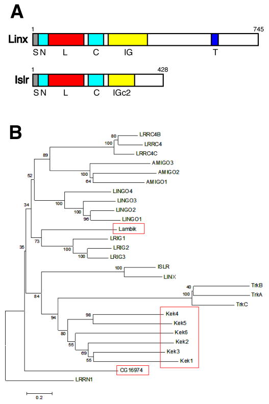
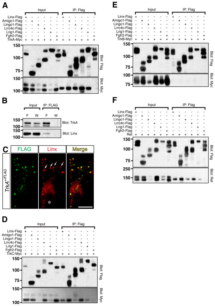
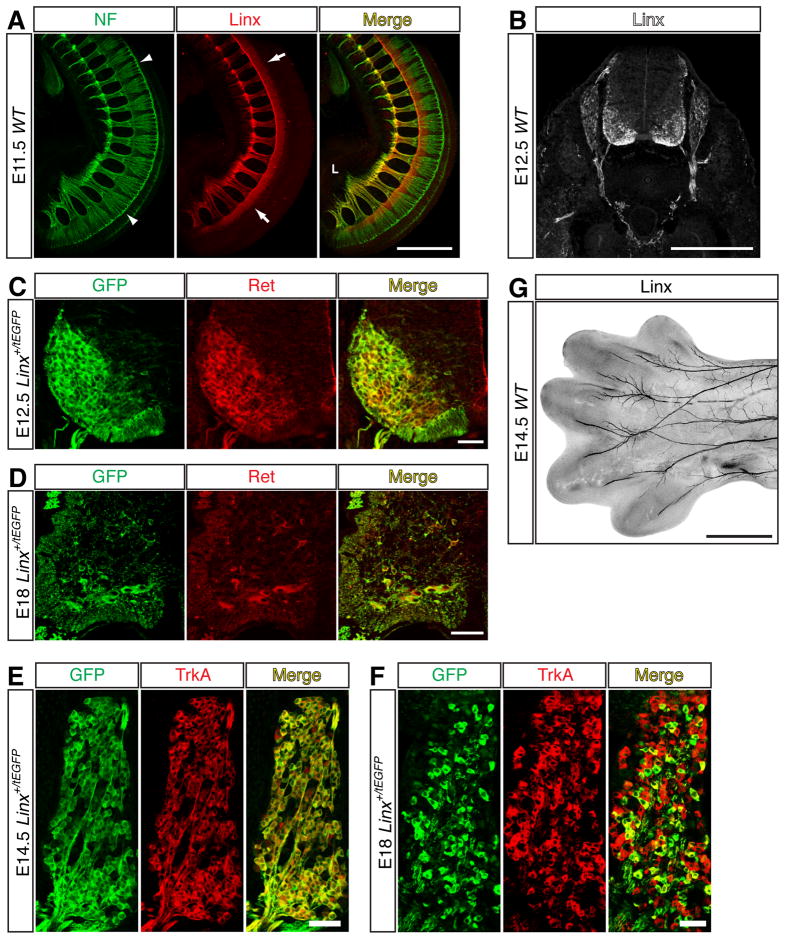
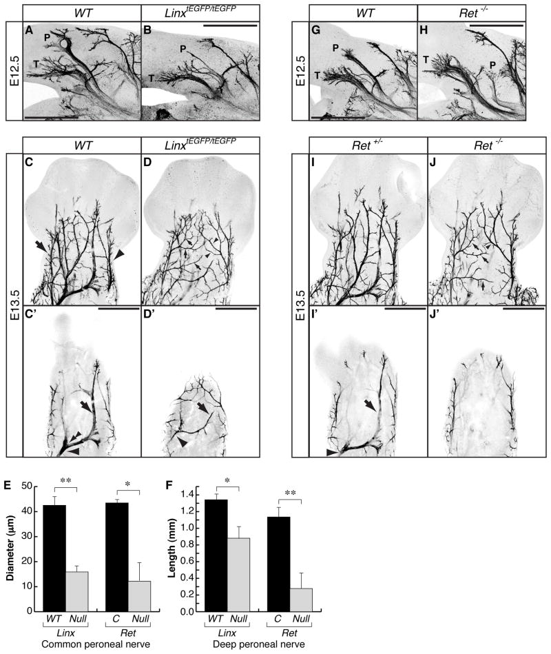
Genome-wide screens were performed to identify transmembrane proteins that mediate axonal growth, guidance and target field innervation of somatosensory neurons. One gene, Linx (alias Islr2), encoding a leucine-rich repeat and immunoglobulin (LIG) family protein, is expressed in a subset of developing sensory and motor neurons. Domain and genomic structures of Linx and other LIG family members suggest that they are evolutionarily related to Trk receptor tyrosine kinases (RTKs). Several LIGs, including Linx, are expressed in subsets of somatosensory and motor neurons, and select members interact with TrkA and Ret RTKs. Moreover, axonal projection defects in mice harboring a null mutation in Linx resemble those in mice lacking Ngf, TrkA, and Ret. In addition, Linx modulates NGF-TrkA- and GDNF-GFRalpha1/Ret-mediated axonal extension in cultured sensory and motor neurons, respectively. These findings show that LIGs physically interact with RTKs and modulate their activities to control axonal extension, guidance and branching.


Figures

Figure 1
Figure 1. Linx Is a LIG Family Member
(A) Domain organizations of Linx and Islr. S: signal peptide, N: leucine rich repeat N-terminal domain, L: leucine-rich repeat domains, C: leucine rich repeat C-terminal domain, IG: immunoglobulin domain, IGc2: immunoglobulin c2-type domain, T: transmembrane domain. (B) Molecular phylogenetic analysis of human and Drosophila LIG family members. The extracellular protein sequences were aligned using the ClustalV software (Higgins et al., 1992). The branch lengths are proportional to the number of amino acid changes. Drosophila LIGs are encircled with red boxes. Note that a rooted tree was generated designating human LRRN1, a more distantly related gene composed of LRRNT, LRR, LRRCT, IGc2 and fibronectin type 3 domains, as an out-group.
Figure 2
Figure 2. Binding of LIG Family Proteins to Trk and Ret Receptor Tyrosine Kinases
(A, D, E, F) Various combinations of recombinant proteins were expressed in 293T cells and subjected to immunoprecipitation experiments using a FLAG antibody. The precipitates were then examined by Western blot analysis to examine interactions with TrkA (A), TrkC (D), TrkB (E) and Ret (F). Protein molecular weight standards (kDa) are shown on the left side of blots. (B) Physical interaction of endogenous Linx and TrkA. Cultured DRG neurons obtained from E13.5 FLAG epitope tagged-TrkA knock-in (TrkAFLAG/FLAG) and wild-type mice were subjected to immunoprecipitation using a FLAG antibody. The precipitates were examined by Western blot analysis to examine interaction. F: TrkAFLAG/FLAG, W: wild-type. (C) Confocal microscopic images of cultured DRG neurons obtained from E13.5 FLAG epitope tagged-TrkA knock-in mice. Endogenous FLAG-TrkA and Linx were detected with FLAG (green) and Linx antibodies (red). Arrows: examples of vesicle-like structures in axons, asterisk: a nucleus of a DRG neuron, bar: 10 μm.
Figure 3
Figure 3. Expression of Linx in Motor and Sensory Neurons
(A) A Z-Stack confocal image for the lateral half of an E11.5 wild-type embryo stained by whole-mount anti-Neurofilament-M (green) and anti-Linx (red) immunostaining. Arrows: ventral spinal cord, arrowheads: dorsal root entry zone, L: lumbar plexus, bar: 0.5 mm. (B) A transverse section of lumbar spinal cord and DRGs of an E12.5 wild-type embryo stained with a Linx antibody. Bar: 0.5 mm. (C and D) Horizontal sections of lumbar spinal cords of Linx+/tEGFP embryos stained with GFP (green) and Ret (red) antibodies at E12.5 (C) and E18 (D). Bars: 50 μm. (E and F) Horizontal sections of lumbar DRGs of Linx+/tEGFP embryos stained with GFP (green) and TrkA (red) antibodies at E14.5 (E) and E18 (F). Bars: 50 μm. (G) A Z-Stack confocal image of a dorsal view of an E14.5 wild-type left hindlimb visualized by whole-mount anti-Linx immunostaining. Bar: 0.5 mm.
Figure 4
Figure 4. Linx Mutant Mice Partially Phenocopy Ret Mutant Mice, and Linx Functions in the GDNF-Ret Signaling Pathway
(A–D) Whole-mount anti-Peripherin immunostaining of left hindlimbs of LinxtEGFP/tEGFP (B, D and D′) and wild-type (A, C and C′) embryos at E12.5 (A and B) and E13.5 (C, C′, D and D′). WT: wild-type, bars: 0.5 mm. (A and B) Z-Stack confocal images of representative posterior views of left hindlimbs taken from 4 LinxtEGFP/tEGFP and wild-type embryos. Note that the dramatic decrease in length and size of peroneal nerve in LinxtEGFP/tEGFP embryos. P: peroneal nerve, T: tibial nerve. (C, C′, D and D′) Z-Stack confocal images of dorsal views of left hindlimbs (C and D) and their raw images depicting common and deep peroneal nerves (C′ and D′). Arrow: sural nerve, arrowhead: saphenous nerve (C). Small arrows: aberrant branches from sural nerve, small arrowheads: aberrant branches from saphenous nerve (D). Arrows: deep peroneal nerves, arrowheads: common peroneal nerves (C′ and D′). Double arrowhead: a point where a common peroneal nerve divides into superficial and deep peroneal nerves (C′) (E) Average diameter of the common peroneal nerve in LinxtEGFP/tEGFP (n=9) compared to wild-type control embryos (n=8) (left), as well as that of Ret/ (n=7) compared to control embryos (n=6) (right). The 6 control embryos for Ret/ in E and F are composed of 2 wild-type and 4 Ret+/. C: control. * and ** indicate p<0.005 and p<5×10⁻⁵, respectively. (F) Average length of the deep peroneal nerve in LinxtEGFP/tEGFP (n=9) compared to wild-type control embryos (n=9) (left), as well as that of Ret−/− (n=7) compared to control embryos (n=6) (right). The length of the deep peroneal nerve was measured from a branching point (double arrowhead shown in C′) to a distal end of the nerve. C: control. * and ** indicate p<0.01 and p<0.005, respectively. (G–J) Whole-mount anti-Peripherin immunostaining of left hindlimbs of Ret−/− (H, J and J′), wild-type (G) and control Ret+/ (I and I′) embryos at E12.5 (G and H) and E13.5 (I, I′, J and J′). Bars: 0.5 mm. (G and H) Z-Stack confocal images of representative posterior views of left hindlimbs of 3 Ret−/− and wild-type embryos. P: peroneal nerve, T: tibial nerve. (I, I′, J and J′) Z-Stack confocal images of dorsal views of left hindlimbs (I and J) and their raw images depicting common and deep peroneal nerves (I′ and J′). Small arrows: aberrant branches from sural nerve, small arrowhead: aberrant branches from saphenous nerve (J). Arrow: deep peroneal nerve, arrowhead: common peroneal nerve (I′).
Figure 5
Figure 5. Functional Interaction between Linx and Ret
(A–E) Confocal images for anti-Peripherin immunostaining of left hindlimbs of wild-type (A), Ret+/− (B), LinxtEGFP/tEGFP (C), LinxtEGFP/tEGFP;Ret+/ (D) and LinxtEGFP/tEGFP;Ret−/− (E) embryos at E13.5. WT: wild-type, arrow: deep peroneal nerve, arrowhead: the point where the common peroneal nerve divides into superficial and deep peroneal nerves, double arrowhead: distal end of the deep peroneal nerve branch to the 2nd an 3rd digits, bars: 0.5 mm. Note that peroneal nerves are completely absent in LinxtEGFP/tEGFP;Ret/ (E). (F) Length of the deep peroneal nerve in various mutants. The length of the deep peroneal nerve was measured from the dividing point (arrowhead in A) to the end of the digital branch to the 2nd and 3rd digits (double arrowhead in A). In the case of absence of the peroneal nerves, the value is considered as zero. Statistical analysis was performed using multiple comparison test. The table shows significance of the difference of mean values. * indicates p<0.05. N.S.: not significant, het: heterozygous, n: number of embryos analyzed for each group. (G) Linx is required for GDNF-dependent motor axon extension. Average axon length of cultured lumbar motor neurons obtained from E13.5 LinxtEGFP/tEGFP and Linx+/tEGFP control embryos. Lumbar motor neurons were cultured in growth media containing CNTF (10 ng/ml) and either the presence or absence of GDNF (10 ng/ml) for 24hr and axonal lengths (n=174 to 200 cells for each condition) were measured of GFP+ and Islet1+ neurons. * and ** indicate p<0.0005 and p<1X10⁻¹⁰, respectively.
Figure 6
Figure 6. Linx Mutant Mice Partially Phenocopy Ngf Mutant Mice in Nerve Extension Defects
(A–H) Whole-mount anti-Peripherin immunostaining of left hindlimbs of LinxtEGFP/tEGFP (B, D, F and H) and wild-type (A, C, E and G) embryos at E14.5. WT: wild-type, 1 and 5: the 1st and 5th digit. Bars: 0.5 mm in A, B, E and F; 0.25 mm in C, D, G and H. (A and B) Z-Stack confocal images of a dorsal view of the left hindlimb. Boxes: magnifications are shown in C and D, arrowheads: peroneal nerve branches. (C and D) High-magnification Z-stack confocal images showing digital branches of the deep peroneal nerve in the 2nd and 3rd digits. Arrows: digital branches to the 3rd digits. (E and F) Z-Stack confocal images of a plantar view of the left hindlimb. Boxes: magnifications are shown in G and H. (G and H) High-magnification Z-stack confocal images showing a lateral digital branch of the lateral plantar nerve in the 5th digit. Arrows: lateral digital branches of the lateral plantar nerve in the 5th digit. (I) Average ratio of the length of a digital branch of the deep peroneal nerve in the 3rd digit of LinxtEGFP/tEGFP (n=11) compared to wild-type (n=13) embryos. * indicates p<0.01. (J) Average ratio of the length of a lateral digital branch of the lateral plantar nerve in the 5th digit of LinxtEGFP/tEGFP (n=9) compared to wild-type (n=10) embryos. * indicates p<0.01. (K–R) Whole-mount anti-Peripherin immunostaining of left hindlimbs of Ngf−/−;Bax−/− (L, N, P and R) and Ngf+/−;Bax/ control (K, M, O and Q) embryos at E14.5. 1 and 5: the 1st and 5th digit. Bars: 0.5 mm in K, L, O and P; 0.25 mm in M, N, Q and R. (K and L) Z-Stack confocal images of a dorsal view of the left hindlimb. Boxes: magnifications are shown in M and N, arrowheads: peroneal nerve branches. Note that peroneal nerve branches are formed in the Ngf−/−;Bax−/− embryo as the control embryo. (M and N) High-magnification Z-stack confocal images showing digital branches of the deep peroneal nerve in the 2nd and 3rd digits. Arrows: digital branches to the 3rd digit. (O and P) Z-Stack confocal images of a plantar view of the left hindlimb. Boxes: magnifications are shown in Q and R. (Q and R) High-magnification Z-stack confocal images showing a lateral digital branch of the lateral plantar nerve in the 5th digit. Arrows: lateral digital branches of the lateral plantar nerve in the 5th digit. (S) Average ratio of the length of a digital branch of the deep peroneal nerve in the 3rd digit of Ngf−/−;Bax−/− (n=7) compared to control (n=7) embryos. Control embryos in S and T are composed of 2 Bax−/−, 1 Bax+/−, 2 Ngf+/−;Bax−/− and 2 wild-type. DN: Ngf −/−;Bax−/−. ** indicates p<0.005. (T) Average ratio of the length of a lateral digital branch of the lateral plantar nerve in the 5th digit of Ngf−/−;Bax−/− (n=7) compared to control (n=7) embryos. DN: Ngf−/−;Bax−/−. *** indicates p<0.0005.
Figure 7
Figure 7. Linx Mutant Mice Partially Phenocopy Ngf Mutant Mice in Nerve Branching Defects, and Linx Functions in the NGF Signaling Pathway
(A–D) Whole-mount anti-Peripherin immunostaining of left hindlimbs of LinxtEGFP/tEGFP (B and D) and Linx+/tEGFP control (A and C) embryos at E15.5. Bars: 0.625 mm in A and B; 0.25 mm in C and D. (A and B) Z-Stack confocal images of a plantar view of the left hindlimb. Boxes: magnifications are shown in C and D, 1 and 5: the 1st and 5th digit. (C and D) High-magnification Z-stack confocal images showing a medial digital branch of the lateral plantar nerve in the 5th digit. Dotted rectangles: medial digital branches of the lateral plantar nerve in the 5th digit, arrows: representative branches with higher-ordered branches. (E and F) Density of branches (E) and density of branches with higher-ordered branches (F) in the medial digital branch of the lateral plantar nerve of the 5th digit in LinxtEGFP/tEGFP (n=12) compared to control (Linx+/tEGFP: n=6; wild-type: n=3) embryos. ** and *** indicates p<0.005 and p<0.001, respectively. (G–J) Whole-mount anti-Peripherin immunostaining of left hindlimbs of Ngf/;Bax/ (H and J) and Bax−/− control (G and I) embryos at E15.5. Bars: 0.625 mm in G and H; 0.25 mm in I and J. (G and H) Z-Stack confocal images of a plantar view of the left hindlimb. Boxes: magnifications are shown in I and J, 1 and 5: the 1st and 5th digit. (I and J) High-magnification Z-stack confocal images showing a medial digital branch of the lateral plantar nerve in the 5th digit. Dotted rectangles: medial digital branches of the lateral plantar nerve in the 5th digit, arrows: representative branches with higher-ordered branches. (K) Density of branches with higher-ordered branches in the medial digital branch of the lateral plantar nerve in the 5th digit of Ngf/;Bax/ (n=4) compared to control (Bax−/−: n=2; Bax+/: n=1; Ngf+/;Bax+/: n=1) embryos. DN: Ngf/;Bax/. * indicates p<0.05. (L) Linx is required for maximal sensitivity to NGF in cultured DRG sensory neurons. Average axon length of cultured DRG sensory neurons obtained from E13.5 LinxtEGFP/tEGFP and Linx+/tEGFP control embryos. DRG sensory neurons were grown in the presence of 0, 3, 9 and 27 ng/ml NGF with a caspase inhibitor, BAF, for 24 hr and axon lengths (n=40 to 47 cells for each condition) were measured for GFP+ neurons using Neurofilament-M staining. *1, *2, *3 and *4 indicates p<0.01, p<0.005, p<0.001 and p<5×10⁻⁷, respectively. (M–O) Phosphorylation of TrkA, Erk and Akt in cultured DRG sensory neurons obtained from E13.5 LinxtEGFP/tEGFP and wild-type embryos. DRG sensory neurons were stimulated with NGF (10 ng/ml) for 5 min and phosphorylation of TrkA Y490 (M), Erk (N) and Akt (O) were examined by Western blot analysis. Immunoblots were then reprobed with a class III β-Tublin (Tuj1) or TrkA antibody. The bands for phosphorylated proteins from 4 independent experiments were measured by densitometry, and fold changes of the band intensity in LinxtEGFP/tEGFP were calculated against that in wild-type after normalizing with the signal intensities of Tuj1 for pErk and pAkt blots or TrkA for a pTrkA blot. WT: wild-type, N.S.: not significant. * indicates p<0.01.
Figure 8
Figure 8. Expression of LIG Family Members in the DRG and Ventral Spinal Cord
(A and C) Double-label in situ hybridization of Linx, Lingo1, Lrrc4b and Amigo1 (red) with TrkA (A) or Ret (C) (green). E13.5 DRG and spinal cord sections were hybridized with indicated cRNA probes. Bars, 100 μm. (B) NGF-dependent induction of LIG family members. The expression of the indicated genes was evaluated by qRT-PCR. Total RNAs were obtained from three independent sets of E13.5 explant DRG cultures grown in the presence or absence of NGF (25 ng/ml). The extent of the gene induction by NGF was evaluated and reported as fold change (>1). * indicates p<0.05 using a paired t-test.

Similar articles

Cited by

References

    1. Airaksinen MS, Koltzenburg M, Lewin GR, Masu Y, Helbig C, Wolf E, Brem G, Toyka KV, Thoenen H, Meyer M. Specific subtypes of cutaneous mechanoreceptors require neurotrophin-3 following peripheral target innervation. Neuron. 1996;16:287–295. - PubMed
    1. Airaksinen MS, Saarma M. The GDNF family: signalling, biological functions and therapeutic value. Nat Rev Neurosci. 2002;3:383–394. - PubMed
    1. Akopian AN, Sivilotti L, Wood JN. A tetrodotoxin-resistant voltage-gated sodium channel expressed by sensory neurons. Nature. 1996;379:257–262. - PubMed
    1. Baloh RH, Enomoto H, Johnson EM, Jr, Milbrandt J. The GDNF family ligands and receptors - implications for neural development. Curr Opin Neurobiol. 2000;10:103–110. - PubMed
    1. Bennett DL, Michael GJ, Ramachandran N, Munson JB, Averill S, Yan Q, McMahon SB, Priestley JV. A distinct subgroup of small DRG cells express GDNF receptor components and GDNF is protective for these neurons after nerve injury. J Neurosci. 1998;18:3059–3072. - PMC - PubMed

Publication types

MeSH terms