. 2009 Oct 15;122(Pt 20):3684-93.
doi: 10.1242/jcs.045674. Epub 2009 Sep 15.

Galectin-3 promotes lamellipodia formation in epithelial cells by interacting with complex N-glycans on alpha3beta1 integrin

Affiliations

Galectin-3 promotes lamellipodia formation in epithelial cells by interacting with complex N-glycans on alpha3beta1 integrin

Chandrassegar Saravanan et al. J Cell Sci. .

Abstract

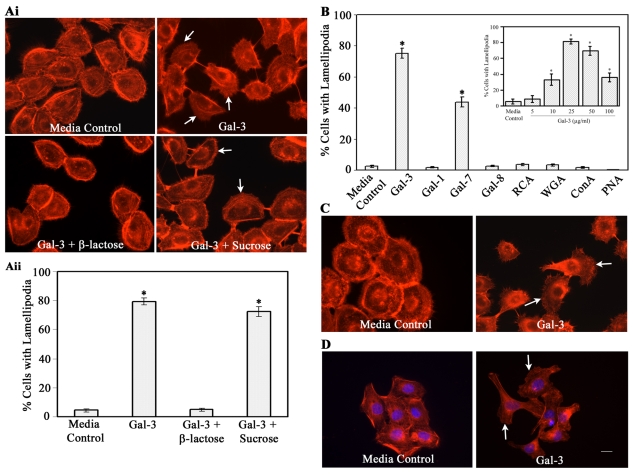
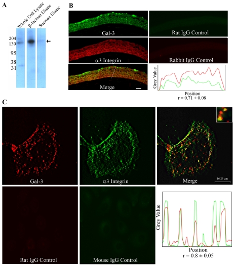
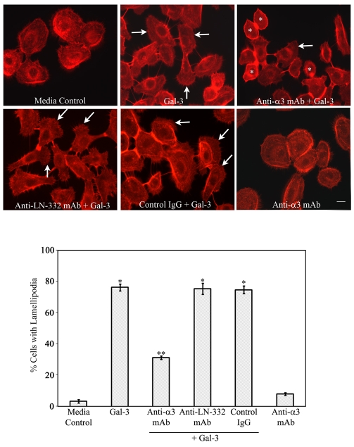
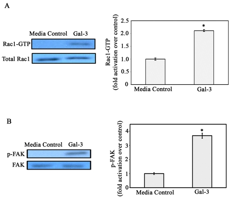
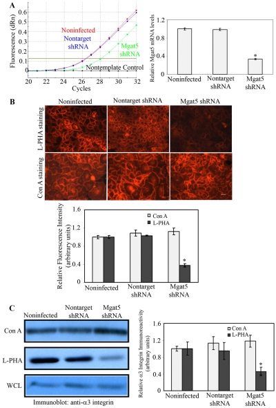
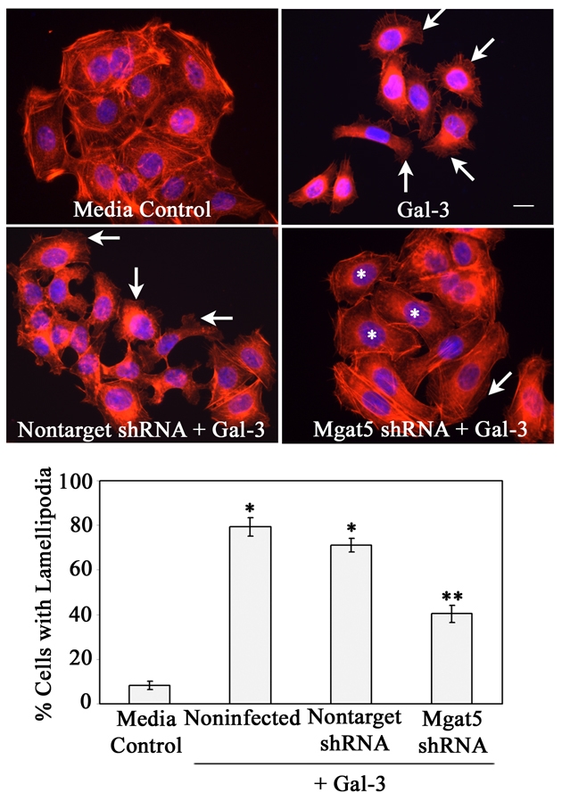
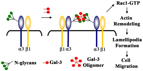
Recent studies have shown that galectin-3 (Gal-3; also known as LGALS3), a beta-galactoside-binding lectin, promotes cell migration during re-epithelialization of corneal wounds. The goal of this study was to characterize the molecular mechanism by which Gal-3 stimulates cell migration. We demonstrate here that exogenous Gal-3, but not Gal-1 or Gal-8, promotes cell scattering and formation of lamellipodia in human corneal epithelial cells in a beta-lactose-inhibitable manner. alpha3beta1 integrin was identified as the major Gal-3-binding protein in corneal epithelial cells by affinity chromatography of cell lysates on a Gal-3-Sepharose column. Preincubation of cells with anti-alpha3 integrin function-blocking antibody significantly inhibited the induction of lamellipodia by Gal-3. Furthermore, exogenous Gal-3 activated both focal adhesion kinase, a key regulator of integrin-dependent intracellular signaling, and Rac1 GTPase, a member of the family of Rho GTPases, well known for its role in the reorganization of the actin cytoskeleton and formation of lamellipodial extensions. Experiments involving knockdown of beta-1,6-N-acetylglucosaminytransferase V, an enzyme that synthesizes high-affinity glycan ligands for Gal-3, revealed that carbohydrate-mediated interaction between Gal-3 and complex N-glycans on alpha3beta1 integrin plays a key role in Gal-3-induced lamellipodia formation. We propose that Gal-3 promotes epithelial cell migration by cross-linking MGAT5-modified complex N-glycans on alpha3beta1 integrin and subsequently activating alpha3beta1-integrin-Rac1 signaling to promote lamellipodia formation.


Figures

Fig. 1.
Fig. 1.
Exogenous Gal-3 and Gal-7, but not Gal-1, Gal-8 or plant lectins, promote formation of lamellipodia in corneal and skin epithelial cells in a carbohydrate-dependent manner. (A) HCLE cells were growth-factor-starved for 2 hours and were then incubated in serum-free medium in the presence or absence of exogenous Gal-3 (25 μg/ml) and saccharides (0.1 M) for 30 minutes. At the end of the incubation period, cells were stained with TRITC-phalloidin and were evaluated using a Leica Optigrid confocal microscope for the presence of lamellipodia (Ai). At least 250 cells were counted from several nonoverlapping microscopic fields and the percentage of cells with lamellipodia was estimated (Aii). No significant lamellipodial protrusions were detected in cells incubated in medium alone. By contrast, the majority of the cells incubated in the presence of Gal-3 had lamellipodial protrusions. The stimulatory effect of exogenous Gal-3 was inhibited by a competing saccharide, β-lactose, but not by a noncompeting disaccharide, sucrose. Data are expressed as mean ± s.e.m. (n=250 cells/group). This experiment was repeated at least five times with reproducible results (*P<0.01 compared with the control). (B) Quantification of fluorescence microscopy data showing that Gal-3 and Gal-7, but not Gal-1, Gal-8 or plant lectins, promote lamellipodia formation in HCLE cells. Inset: Cells were incubated with varying concentrations of Gal-3. (C,D) Gal-3 also promotes lamellipodia formation in primary cultures of human corneal (C) and skin epithelial (D) cells. White arrows indicate lamellipodia. Scale bar: 16 μm.
Fig. 2.
Fig. 2.
(A) α3 Integrin is a major Gal-3-binding protein. HCLE cell lysate was applied to a Gal-3 affinity column, the column was first eluted with a noncompeting disaccharide, sucrose, and then with a competing sugar, β-lactose. Unfractionated extract (2.5 μg protein) and the glycoproteins eluted from the Gal-3-affinity column (derived from 200 μg unfractionated extract) were resolved in reducing 10% SDS-PAGE gels and the protein blots of the gels were processed for immunostaining using a rabbit anti-human α3 integrin polyclonal antibody. Note that a major antibody-reactive 130-kDa glycoprotein (arrow) was detected in the β-lactose but not in the sucrose eluate. (B) α3 integrin and Gal-3 are colocalized in mouse corneal epithelium. Frozen sections (10 μm thick) of mouse corneas were fixed with ice-cold methanol and co-labeled with anti-human Gal-3 rat monoclonal and anti-human α3 integrin rabbit polyclonal antibodies. Gal-3 is green and α3 integrin is red. The merged image shows complete colocalization of Gal-3 and α3 integrin at both cell-cell and cell-matrix junctions. (Bottom right) The intensity plot along the line drawn in the merged image showing a high Pearson's correlation coefficients (r=0.71±0.08; n=5) indicative of substantial Gal-3 (green) colocalization with α3 integrin (red). Scale bar: 20 μm. (C) Colocalization of Gal-3 and α3 integrin in epithelial cells in culture. The HCLE cells were exposed to Gal-3 and were processed for immunostaining using Gal-3 and α3-integrin-specific monoclonal antibodies. A single section of the z-stack representing the bottom of the cell shows that Gal-3 (red) and α3 integrin (green) colocalize at cell-matrix interaction sites specifically in lamellae (base of lamellipodia) and at the rear of the migrating cell (merged image). The inset is an enlarged image of the boxed region showing discrete yellow spots indicating the colocalization of the lectin and integrin. (Bottom right) The intensity plot along the line drawn in the merged image showing a high Pearson's correlation coefficient (r=0.8±0.05; n=5) suggestive of significant colocalization of Gal-3 (red) with α3 integrin (green). Note that no staining was detected in the control samples treated with rat or mouse IgG.
Fig. 3.
Fig. 3.
An anti-α3 integrin but not anti-LN-332 function-blocking mAb inhibits Gal-3-induced lamellipodia formation. HCLE cells were plated on chamber slides and were incubated with 10 μg/ml of the anti-α3 integrin mAb (P1B5), anti-LN-332 mAb (P3H9-2) or a control mAb (anti-α2 integrin, Clone P1E6) for 30 minutes, prior to exposure to Gal-3. At the end of the incubation period, the cells were evaluated for the presence of lamellipodial protrusions as described in Fig. 1 legend (bottom panel). Note that preincubation with anti-α3 integrin mAb blocked Gal-3-induced lamellipodia formation in the majority of the cells (* in the anti-α3 integrin + Gal-3 panel). By contrast, preincubation of cells with anti-LN-332 mAb, or anti-α2 integrin mAb or control IgG, did not prevent Gal-3-induced lamellipodial protrusions (IgG + Gal-3 panel). The cells incubated in medium alone and medium containing Gal-3 served as negative and positive controls, respectively. Data are expressed as mean ± s.e.m. (n=250 cells/group). This experiment was repeated at least three times with reproducible results (**P<0.01 compared with Gal-3-treated cells; *P<0.01 compared with control). White arrows indicate lamellipodia. Scale bar: 16 μm.
Fig. 4.
Fig. 4.
Gal-3 activates Rac1 GTPase and FAK in corneal epithelial cells. (A) HCLE cells were growth-factor-starved and were incubated in the presence or absence of Gal-3 in serum-free medium. Cell extracts were subjected to pulldown assays using GST-PAK to examine GTP-bound Rac1 (active Rac1) levels. Precipitated GTP-Rac1 as well as total cell lysates were examined by immunoblot analysis using a mouse anti-Rac1 mAb. The intensity of each band was quantified by densitometry and the ratios of GTP-loaded Rac1 to total Rac1 were determined and normalized to cells exposed to serum-free medium alone (media control). Note that the levels of total Rac1 (GTP-bound + GDP-bound forms) were similar regardless of whether the cells were exposed to Gal-3 or not. By contrast, within 30 seconds of exposure to Gal-3, there was a 2.3-fold increase in the amount of activated Rac1 (bar graph), and enhanced Rac1 activity was maintained up to 10 minutes (data not shown). Values are mean ± s.e.m. of three separate experiments. *P<0.01 compared to media control. (B) Electrophoresis blots were probed with anti-FAK or anti-Y397 phosphorylated FAK monoclonal antibodies. Note that FAK is activated (p-FAK lanes) after exposure to Gal-3, whereas total FAK levels (FAK lanes) are similar in the control and Gal-3-exposed cells. The intensity of each band was quantified and ratios of p-FAK to total FAK was calculated and normalized to control values (bar graph). *P<0.05 when compared with control; data are mean ± s.e.m. from three independent experiments.
Fig. 5.
Fig. 5.
Targeted knockdown of MGAT5 by shRNA reduces β1,6GlcNAc-branched N-glycans of α3 integrin in epithelial cells. (A) RT-qPCR. Total RNA was isolated from noninfected control, MGAT5 shRNA- and nontarget shRNA-expressing cells. (Left) RT-qPCR was performed using primers specific for human MGAT5. Left panel: original amplification plots of MGAT5 mRNAs showing significantly lower signals in cells expressing MGAT5 shRNA compared with cells expressing nontarget shRNA, and noninfected control cells. (Right) The data were normalized against GAPDH and the fold change was calculated using noninfected control cells as a calibrator (n=3). *P<0.05 compared with noninfected or nontarget shRNA-expressing cells. (B) Lectin staining. Noninfected, nontarget shRNA-, and MGAT5 shRNA-expressing cells grown on chamber slides were fixed with 4% paraformaldehyde and then stained with either rhodamine-conjugated L-PHA (specificity: core β1,6GlcNAc-branched N-glycans) or Con A (specificity: high mannose and bisected hybrid N-glycans). The staining intensity was quantified from 10 images in each experimental condition and normalized to that of noninfected cells (bar graph). Top panel shows representative micrographs of stained cells. Note that the binding of L-PHA but not Con A was reduced significantly in cells expressing MGAT5 shRNA. Data are expressed as mean ± s.e.m. The experiment was repeated three times with reproducible results. *P<0.05 compared with noninfected and nontarget shRNA-expressing cells. Scale bar: 32 μm. (C) Western blotting. To determine whether knockdown of MGAT5 expression reduces β1,6GlcNAc-branched complex N-glycans on α3β1 integrin, RIPA buffer extracts from confluent cultures of noninfected, MGAT5 shRNA-, and nontarget shRNA-expressing cells (600 μg total protein each) were incubated with agarose-bound Con A or L-PHA (1 hour, 4°C). Following incubation, the beads were washed, boiled in SDS-PAGE sample buffer (without mercaptoethanol) to release bound proteins, centrifuged, and supernatants were electrophoresed in SDS polyacrylamide gels. Samples derived from 2.5 μg original cell protein were electrophoresed in each lane. Protein blots of the gels were then processed for immunostaining with rabbit anti-α3 integrin. Note that an equivalent level of anti-α3-integrin-reactive component is present in the Con-A-bound fraction and whole cell extract (WCL). By contrast, significantly reduced level of anti-α3-integrin-reactive component was detected in the L-PHA-bound fraction in MGAT5 knockdown cells. Also, no anti-α3-integrin-reactive components were detected in the supernatants of cell extracts incubated with agarose beads alone or in the blots of cell extracts not exposed to primary antibody (not shown). Relative binding of lectins to α3 integrin was calculated by densitometric analysis (bar graph). Values are mean ± s.e.m. of three independent experiments. *P<0.05 compared with noninfected and nontarget shRNA-expressing cells.
Fig. 6.
Fig. 6.
MGAT5 knockdown inhibits Gal-3-induced lamellipodia formation in epithelial cells. Epithelial cells expressing either nontarget shRNA or MGAT5 shRNA or noninfected cells were exposed to serum-free medium in the presence and absence of Gal-3 for 30 minutes. At the end of the incubation period, cells were stained with TRITC-phalloidin (red) and DAPI (blue) and were then evaluated for the presence of lamellipodial protrusions, under a fluorescent microscope. Representative images are shown in the top panel. The majority of cells incubated with Gal-3, but not with media alone, show lamellipodial protrusions (arrows). Note that the stimulatory effect of Gal-3 on lamellipodia formation was inhibited in MGAT5 shRNA-expressing cells (* in MGAT5 shRNA+Gal-3 panel), but not in the cells expressing nontarget shRNA. The percentage of cells with lamellipodial protrusions were calculated after examination of at least 250 cells from each experimental sample (bar graph). Data are expressed as mean ± s.e.m. (n=250 cells/group). The experiment was repeated three times with reproducible results. *P<0.01 compared with control; **P<0.01 compared with noninfected cells exposed to Gal-3. Scale bar: 16 μm.
Fig. 7.
Fig. 7.
Proposed model of Gal-3-mediated signaling in epithelial cells leading to the formation of lamellipodial protrusions and cell migration. According to this model, Gal-3, by virtue of its multivalency (Fred Brewer, 2002), cross-links and clusters α3β1 integrin on the cell surface at the leading edge of the migrating epithelium. The clustering of α3β1 integrin activates FAK and Rac1 and this, in turn, promotes lamellipodia formation, cell migration and re-epithelialization of wounds. This model is suggested based on our findings that: (1) α3β1 integrin is a major Gal-3-binding partner and a function-blocking anti-α3 integrin mAb blocks the Gal-3-mediated lamellipodia formation, (2) Gal-3 interacts with MGAT5-modified complex N-glycans on α3β1 integrin, (3) Gal-3 activates Rac1, a member of Rho GTPases, and the published findings showing that: (i) Gal-3 cross-links cell surface receptors (e.g. EGF and TGFβ receptors) by interacting with MGAT5-modified complex N-glycans to promote their signal transduction (Partridge et al., 2004), and (ii) α3β1-integrin–Rac1 signaling is essential to promote lamellipodia formation (Choma et al., 2004).

References

    1. Adams, J. C. (2001). Cell-matrix contact structures. Cell Mol. Life Sci. 58, 371-392. - PMC - PubMed
    1. Bao, Q. and Hughes, R. C. (1999). Galectin-3 and polarized growth within collagen gels of wild-type and ricin-resistant MDCK renal epithelial cells. Glycobiology 9, 489-495. - PubMed
    1. Bellis, S. L. (2004). Variant glycosylation: an underappreciated regulatory mechanism for beta1 integrins. Biochim. Biophys. Acta 1663, 52-60. - PubMed
    1. Boukamp, P., Petrussevska, R. T., Breitkreutz, D., Hornung, J., Markham, A. and Fusenig, N. E. (1988). Normal keratinization in a spontaneously immortalized aneuploid human keratinocyte cell line. J. Cell Biol. 106, 761-771. - PMC - PubMed
    1. Brewer, C. F. (2004). Thermodynamic binding studies of galectin-1, Gal-3 and -7. Glycoconj. J. 19, 459-465. - PubMed

Publication types

MeSH terms