

Principles, techniques, and applications of T2*-based MR imaging and its special applications
- PMID: 19755604
- PMCID: PMC2799958
- DOI: 10.1148/rg.295095034
Principles, techniques, and applications of T2*-based MR imaging and its special applications
Abstract
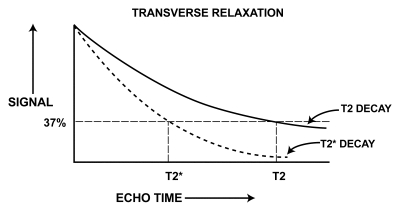
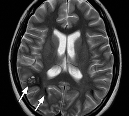
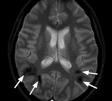
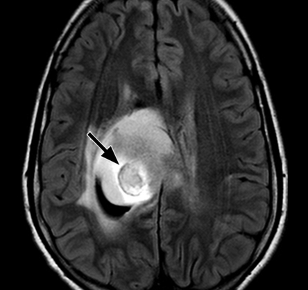
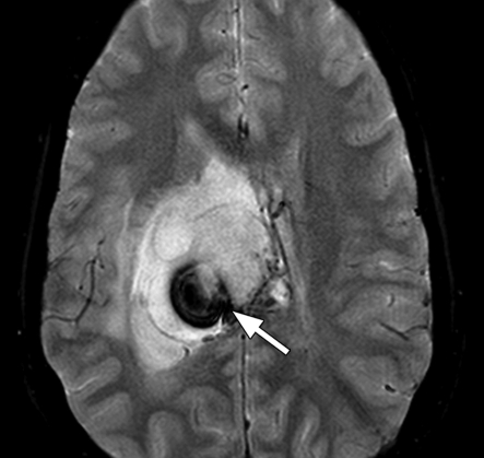
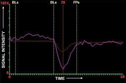
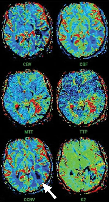
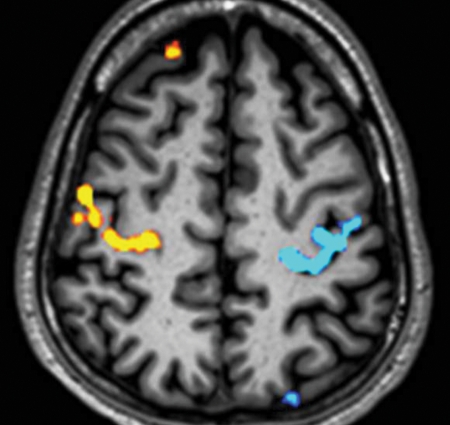
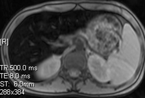
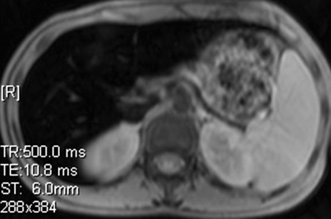
T2* relaxation refers to decay of transverse magnetization caused by a combination of spin-spin relaxation and magnetic field inhomogeneity. T2* relaxation is seen only with gradient-echo (GRE) imaging because transverse relaxation caused by magnetic field inhomogeneities is eliminated by the 180 degrees pulse at spin-echo imaging. T2* relaxation is one of the main determinants of image contrast with GRE sequences and forms the basis for many magnetic resonance (MR) applications, such as susceptibility-weighted (SW) imaging, perfusion MR imaging, and functional MR imaging. GRE sequences can be made predominantly T2* weighted by using a low flip angle, long echo time, and long repetition time. GRE sequences with T2*-based contrast are used to depict hemorrhage, calcification, and iron deposition in various tissues and lesions. SW imaging uses phase information in addition to T2*-based contrast to exploit the magnetic susceptibility differences of the blood and of iron and calcification in various tissues. Perfusion MR imaging exploits the signal intensity decrease that occurs with the passage of a high concentration of gadopentetate dimeglumine through the microvasculature. Change in oxygen saturation during specific tasks changes the local T2*, which leads to the blood oxygen level-dependent effect seen at functional MR imaging. The basics of T2* relaxation, T2*-weighted sequences, and their clinical applications are presented, followed by the principles, techniques, and clinical uses of four T2*-based applications, including SW imaging, perfusion MR imaging, functional MR imaging, and iron overload imaging.
(c) RSNA, 2009.
Figures


References
-
- Mugler JP., III Basic principles. In: Edelman RR, Hesselink JR, Zlatkin MB, Crues JV, eds. Clinical magnetic resonance imaging 3rd ed Philadelphia, Pa: Saunders Elsevier, 2006; 23–57
-
- Nitz WR, Reimer P. Contrast mechanisms in MR imaging. Eur Radiol 1999;9:1032–1046 - PubMed
-
- Hendrick RE. Image contrast and noise. In: Stark DD, Bradley WG, eds. Magnetic resonance imaging 3rd ed St Louis, Mo: Mosby, 1999; 43–68
-
- Haacke EM, Tkach JA, Parrish TB. Reduction of T2* dephasing in gradient field-echo imaging. Radiology 1989;170:457–462 - PubMed
-
- Frahm J, Haenicke W. Rapid scan techniques. In: Stark DD, Bradley WG, eds. Magnetic resonance imaging 3rd ed St Louis, Mo: Mosby, 1999; 87–124
Publication types
MeSH terms
Grants and funding
LinkOut - more resources
Full Text Sources
Other Literature Sources
Medical
