. 2009 Sep 15;2(88):ra55.
doi: 10.1126/scisignal.2000304.

Differential interactions of FGFs with heparan sulfate control gradient formation and branching morphogenesis

Affiliations

Differential interactions of FGFs with heparan sulfate control gradient formation and branching morphogenesis

Helen P Makarenkova et al. Sci Signal. .

Abstract

The developmental activities of morphogens depend on the gradients that they form in the extracellular matrix. Here, we show that differences in the binding of fibroblast growth factor 7 (FGF7) and FGF10 to heparan sulfate (HS) underlie the formation of different gradients that dictate distinct activities during branching morphogenesis. Reducing the binding affinity of FGF10 for HS by mutating a single residue in its HS-binding pocket converted FGF10 into a functional mimic of FGF7 with respect to gradient formation and regulation of branching morphogenesis. In particular, the mutant form of FGF10 caused lacrimal and salivary gland epithelium buds to branch rather than to elongate. In contrast, mutations that reduced the affinity of the FGF10 for its receptor affected the extent, but not the nature, of the response. Our data may provide a general model for understanding how binding to HS regulates other morphogenetic gradients.


Figures
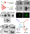
Fig. 1
Fig. 1
FGF gradients can regulate the morphogenesis of LG buds. (A, B) A hypothetical model of the regulation of branching morphogenesis by FGFs with different affinities for HS. FGFs applied locally (asterisk) diffuse differently depending on their affinities for HS. (A) An FGF with a high affinity for HS has restricted diffusion (red sector) through the ECM and signals only to those cells (red crosses) that are most proximal to the FGF source resulting in elongation of the bud towards the source. (B) An FGF with low affinity for HS has a much broader diffusion range, which leads to proliferation of cells that are proximal and distal to the FGF source, which results in branching. (C to F) Effects of FGF10 and FGF7 on the growth and migration of the isolated lacrimal bud. Within 24 hours of exposure, buds adjacent to an FGF10-loaded bead elongated towards the bead (C, D), whereas buds adjacent to an FGF7-loaded bead branched instead (E, F) (five independent experiment were performed: 8–10 explants per condition per experiment). (G to J) Gradient formation by, and response of isolated lacrimal buds to, an FGF10-loaded bead in collagen gel in the absence (G and I) or presence (H and J) of soluble heparin. The extended FGF gradient (H) induced branching (J) instead of elongation (five experiments; 8–10 explants per condition per experiment). (K) Four FGF10-loaded beads placed around an LG epithelial bud induced branching (three experiments; 24 explants). (L) Schematic describing an experiment in which LG buds are briefly exposed to soluble FGF10 (red) or BSA (blue) prior to culture near an FGF10-loaded bead. (M to P) Short exposure of whole lacrimal buds (distal and proximal parts) to FGF10 (25 ng/ml) induced branching near the FGF10-loaded bead (L, O, and P), whereas buds treated with BSA migrated towards the bead (L, M, and N) (seven experiments performed; 8–10 explants per condition per experiment).
Fig. 2
Fig. 2
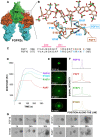
Mutations in the HS-binding domain of FGF10 change its affinity for HS, diffusion profile, and biological function. (A) The structure of the FGF2-FGFR2c-heparin ternary complex is shown and depicted as a surface. FGF2 is colored orange, D2 of FGFR2c is colored green, D3 of FGFR2c is colored cyan, and an 8-mer heparin molecule is colored red. The box indicates the region that is expanded and shown in (B), in which FGF7 and FGF10 are superimposed on the structure of FGF2; FGF2 itself is not shown. The heparin molecule has been borrowed from the FGF2-FGFR2c-heparin complex to show its interactions with FGF7 and FGF10. The FGF7 carbon backbone is colored orange, and the amino acids changes that were introduced into FGF10 have been labeled in orange. The carbon backbone of FGF10 is colored cyan, and the residues that were mutated have been labeled in cyan. The carbon backbone of heparin is colored tan. In FGF7, FGF10, and heparin, oxygen atoms are depicted as red, nitrogen atoms as blue, and sulfur atoms as green. (C) Sequence alignment of the heparin-binding sites of human FGF7 and FGF10. The heparin binding residues in FGF7 and FGF10 are colored in orange and cyan, respectively. (D) SPR analysis of the interaction of FGFs with heparin. Representative sensograms of injections of 40 nM of a given FGF over a streptavidin chip with immobilized biotinylated heparin are shown: FGF7 (red), FGF10 (blue), FGF10R193K (cyan), FGF10R187V (green). The biosensor chip response is indicated in Response Units (y-axis) as a function of time (x-axis) (five experiments was performed). (E to F) The difference in diffusion profiles of FGF10, FGF7, and FGF10-HS mutants. FGF10 formed a sharp gradient around the bead, whereas FGF7 and FGF10R187V diffused more freely through the matrix. FGF10R193K exhibited slightly increased diffusion compared to that of FGF10. (G) Biological responses of LG epithelial buds exposed to FGF7, FGF10, or FGF10-HS mutants on acrylic beads. FGF10R187V and FGF7 induced the same biological response in the bud (mainly branching), whereas FGF10 induced elongation of the bud. FGF10R193K had an intermediate effect; it induced both branching and elongation of the bud (15 experiments were performed).
Fig. 3
Fig. 3
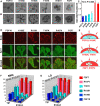
Biological responses of isolated LG and SMG epithelial explants to FGF7, FGF10, and FGF10-HS mutants in an ECM-diffusion assay. Growth responses of epithelial explants of LG (A) or SMG (B). (C) The morphology of the LG explant (the ratio between bud width and length) correlated with the difference in HS affinity and gradient formation of the FGF. The ratio of the width to the length of epithelial buds was determined in four independent experiments (5 to 7 explants of each kind); the Student's t test was used for statistical analysis. (D) BrdU labeling (red) of SMG explants grown in the presence of FGFs. An antibody specific for syndecan-1 (green) was used to label all of the epithelial cells. Explants exposed to the sharp gradients produced by the restricted diffusion of FGF10 and FGF10K195E showed proliferating cells only at the bud tips (which are near the interface between the gel and the medium). In contrast, FGF10R193K and FGF10T197K induced cell proliferation at the tips and also more distally in the explants. Both FGF7 and FGF10R187V, which could diffuse freely throughout the gel, induced cell proliferation throughout the whole explant. (E) Different FGFs permeating into the gel are differentially captured by HS (indicated schematically in red) and are thus likely to form different gradients that elicit different cellular responses. Quantification of BrdU labeling in SMG (F) and LG (G) explants exposed to FGF ligands. Explants exposed to FGF10R187V showed the same labeling profile as those that were exposed to FGF7. BrdU quantification for LG and SMG explants was performed in four independent experiments (6 to 8 explants of each kind).
Fig. 4
Fig. 4
Receptor-binding mutants of FGF10 induced less elongation of LG and SMG epithelial explants than did WT-FGF10, but did not induce a switch from an elongation response to a branching response. Gel-embedded explants were exposed either to FGFs loaded on a bead (A) or to FGFs added to the media in a gel permeation assay (B). The mutant FGF10 with the strongest impact on the strength or duration of the interaction between FGF10 and FGFR2b completely blocked epithelial growth.
Fig. 5
Fig. 5
Digestion of HSGAG in the ECM or competition of FGF10 binding to the ECM with heparin changed the biological response of SMG epithelial explants to FGF10. SMG epithelia were cultured in a 3D laminin gel with FGF10 for 44 h. The 3D matrix was either pretreated with chondroitinase ABC (ChABC) or heparitinase (both at 20 U/ml) and then washed extensively with media to remove the digested GAGs and enzymes. Other groups had exogenous heparitinase (20 U/ml) or heparin (0.5 μg/ml) added to the media, with no washout of GAGs, and one group was cultured with FGF7 alone ** p < 0.01 as measured by ANOVA.
Fig. 6
Fig. 6
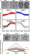
FGF10R187V and FGF7 induced nearly identical gene expression profiles. (A and B) FGF7 and FGF10R187V produced similar epithelial morphologies in the SMG (A) and a similar gene expression profile (B), whereas FGF10R193K induced responses more similar to those elicited by FGF10 (A and B). K-means cluster analysis of changes in epithelial gene expression that were detected with an Agilent Mouse Genome Microarray after 44 hrs of culture with FGF10, FGF10R193K, FGF10R187V, or FGF7. (C and D) FGF7 and FGF10R187V induced similar epithelial morphologies in the LG (C) and a similar gene expression profile (D). Gene expression was examined with a PCR-array focused on the MAPK signaling pathway and the expression profiles of all groups were compared to that of FGF7. Genes were clustered into 3 groups: genes that showed a greater than 2-fold increase in expression in response to FGF10 relative to that of FGF7 or FGF10R187V (red), genes that showed at least a 2-fold decrease in expression in response to FGF10 relative to that of FGF7 or FGF10R187V (green), and genes that showed a less than 2-fold change in expression in either direction (gray).

References

    1. Mohammadi M, Olsen SK, Goetz R. A protein canyon in the FGF-FGF receptor dimer selects from an a la carte menu of heparan sulfate motifs. Curr Opin Struct Biol. 2005;15:506–516. - PubMed
    1. Mohammadi M, Olsen SK, Ibrahimi OA. Structural basis for fibroblast growth factor receptor activation. Cytokine Growth Factor Rev. 2005;16:107–137. - PubMed
    1. Itoh N, Ornitz DM. Functional evolutionary history of the mouse Fgf gene family. Dev Dyn. 2008;237:18–27. - PubMed
    1. Ornitz DM, Itoh N. Fibroblast growth factors. Genome Biol. 2001;2 REVIEWS3005. - PMC - PubMed
    1. Schlessinger J, Plotnikov AN, Ibrahimi OA, Eliseenkova AV, Yeh BK, Yayon A, Linhardt RJ, Mohammadi M. Crystal structure of a ternary FGF-FGFR-heparin complex reveals a dual role for heparin in FGFR binding and dimerization. Mol Cell. 2000;6:743–750. - PubMed

MeSH terms

LinkOut - more resources