Review
. 2009 Oct;7(10):703-14.
doi: 10.1038/nrmicro2218.

The structural biology of type IV secretion systems

Affiliations
Review

The structural biology of type IV secretion systems

Rémi Fronzes et al. Nat Rev Microbiol. 2009 Oct.

Abstract

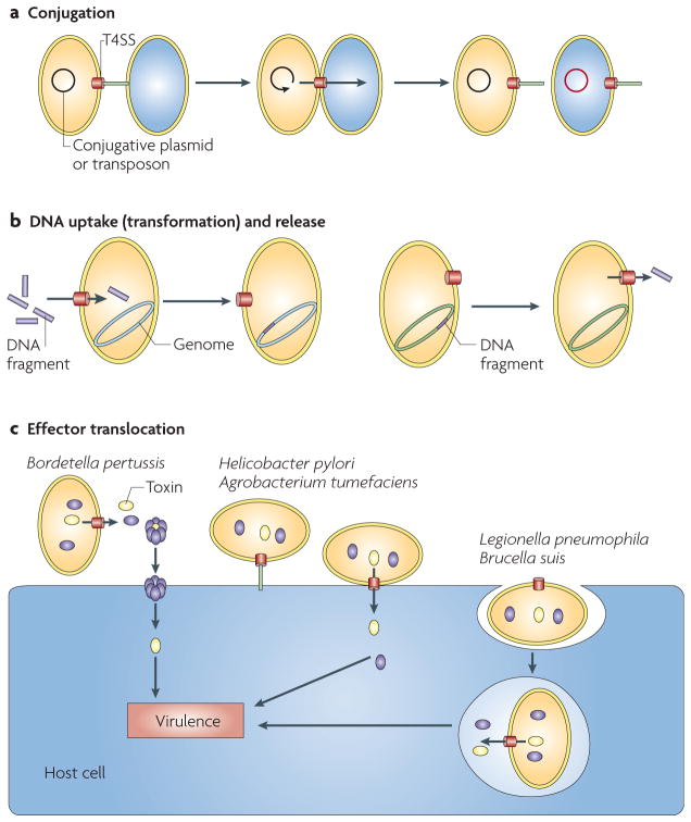
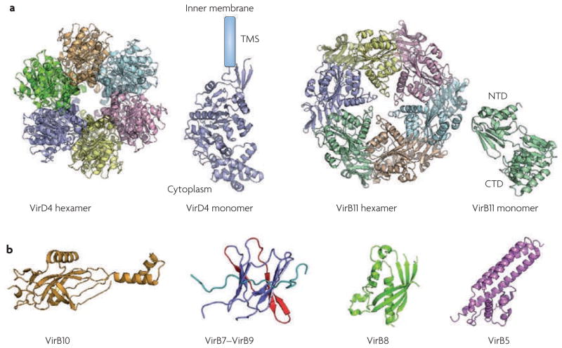
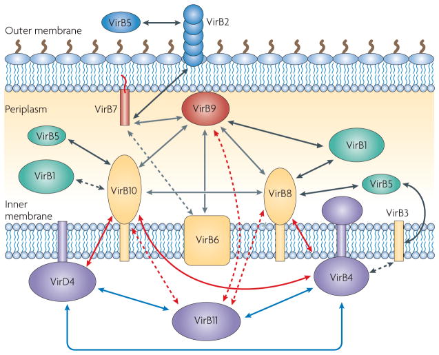
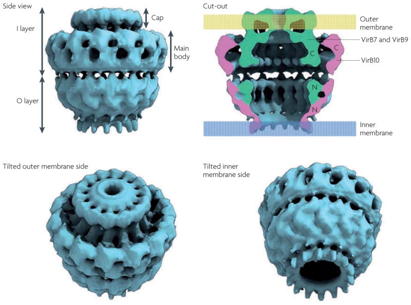
Type IV secretion systems (T4SSs) are versatile secretion systems that are found in both Gram-negative and Gram-positive bacteria and secrete a wide range of substrates, from single proteins to protein-protein and protein-DNA complexes. They usually consist of 12 components that are organized into ATP-powered, double-membrane-spanning complexes. The structures of single soluble components or domains have been solved, but an understanding of how these structures come together has only recently begun to emerge. This Review focuses on the structural advances that have been made over the past 10 years and how the corresponding structural insights have helped to elucidate many of the details of the mechanism of type IV secretion.


Figures

Figure 1
Figure 1. Schematic of the role of type IV secretion in bacteria
The three groups of type IV secretion apparatus are shown. a | Conjugative type IV secretion systems (T4SSs) deliver plasmids or transposons from donor cell to recipient cell in Gram-negative and Gram-positive bacteria. b | DNA uptake (transformation) and release T4SSs mediate the exchange of DNA with the extracellular milieu. c | Effector translocation T4SSs deliver DNA or protein substrates to eukaryotic cells and are directly involved in the virulence of many pathogenic Gram-negative bacteria (for further details, see REFS 10,11,128,129).
Figure 2
Figure 2. Atomic structures of type IV secretion system components or domains
All structures are shown in a ribbon representation. a |The structure of the VirD4 homologue TrwB hexamer and monomer is shown on the left; TrwB is encoded by the Escherichia coli conjugative plasmid R388 (REF. 18). The crystal structures of the Helicobacter pylori VirB11 homologue HP5025 hexamer and monomer are shown on the right (REFS 33,34). b | From left to right, the structures shown are: the crystal structure of the periplasmic carboxy-terminal domain of the H. pylori VirB10 homologue ComB10 (REF. 55); the nuclear magnetic resonance spectroscopy structure of the C-terminal domain of the VirB9 homologue TraO (in blue, with the β-appendage in red), which is encoded by the E. coli pKM101 conjugative plasmid, in complex with the VirB7 homologue TraN (in cyan); the crystal structure of the periplasmic C-terminal domain of VirB8 from Brucella suis and Agrobacterium tumefaciens,; and the crystal structure of the VirB5 homologue TraC, which is encoded by the E. coli conjugative plasmid pKM101 (REF. 102). CTD, carboxy-terminal domain; NTD, amino-terminal domain; TMS, transmembrane segment.
Figure 3
Figure 3. Schematic of the localization of the Agrobacterium tumefaciens VirB/D type IV secretion components and their interactions
The VirD4 and VirB1–VirB11 type IV secretion system (T4SS) components are shown according to their proposed localization (cytoplasmic ATPases are shown in purple, inner membrane proteins in yellow, periplasmic proteins in green, outer membrane proteins in red and extracellular proteins in blue). Interactions between ATPases are shown with blue arrows. Interactions between ATPases and any non-ATPase T4SS component are shown with red arrows. Interactions between T4SS core components are shown with grey arrows. Black arrows indicate other interactions. Possible interactions are indicated by dotted arrows. The publications corresponding to each interaction are: VirB1–VirB8 (REFS 45,73,74); VirB1–VirB9 (REFS 45,74); VirB1–VirB10 (REF. 45); VirB2–VirB5 (REFS 44,100,101,103); VirB2–VirB7 (REFS 70,130); VirB3–VirB4 (REFS 44,85,131,132); VirB3–VirB5 (REF. 133); VirB4–VirB8 (REFS 44,45,48); VirB4–VirB10 (REFS –48); VirB4–VirB11 (REFS 45,49); VirB4–VirD4 (REFS 48,49); VirB5–VirB8 (REF. 44); VirB5–VirB10 (REF. 44); VirB6–VirB7 (REFS 70,71); VirB6–VirB9 (REF. 71); VirB7–VirB9 (REFS ,,,,–139); VirB8–VirB9 (REFS 48,69,70,72); VirB8–VirB10 (REFS 69,70,72); VirB8–VirB11 (REF. 45); VirB9–VirB10 (REFS 48,53,58,69,70,72,140); VirB9–VirB11 (REFS 45,48); VirB10–VirB11 (REF. 45); VirB11–VirD4 (REF. 49).
Figure 4
Figure 4. Structure of a type IV secretion core complex
The core complex is composed of TraN (a VirB7 homologue), TraO (a VirB9 homologue) and TraF (a VirB10 homologue), which are encoded by the Escherichia coli conjugative plasmid pKM101. This structure was obtained using cryoelectron microscopy and is viewed from the side (upper left panel), tilted towards the outer membrane side (lower left panel) and tilted towards the inner membrane side (lower right panel). The cut-out view (upper right panel) details the proposed transmembrane regions and the localization of the VirB7, VirB9 and VirB10 homologues within the structure. C, carboxy-terminal domain; N, amino-terminal domain.
Figure 5
Figure 5. Translocation of T-DNA
The figure shows the T-DNA translocation pathway within the type IV secretion apparatus of Agrobacterium tumefaciens, as determined by the transfer DNA immunoprecipitation asssay. The substrate pathway is shown in red arrows. The T-DNA interacts directly with VirD4, VirB11, VirB6, VirB8 and finally with VirB9–VirB2. The other components influence the transfer of the substrate at different stages, as shown by black arrows.
Figure 6
Figure 6. Schematic of the assembly and function of a type IV secretion system
a | The translocation pore consists of the core complex proteins VirB7, VirB9 and VirB10, the inner-membrane pore proteins VirB6 and VirB8, VirB4, the pilus-associated proteins VirB2, VirB3 and VirB5, and possibly VirB1. b | Depending on unidentified stimuli, the complex could further assemble to secrete a substrate, by the addition of VirD4 and VirB11. Arrows around VirB10 indicate potential conformational changes linked to ATP-binding and hydrolysis by cytoplasmic–inner membrane ATPases. c | Alternatively, the complex could assemble a pilus with the addition of VirB11.

References

    1. Grohmann E, Muth G, Espinosa M. Conjugative plasmid transfer in Gram-positive bacteria. Microbiol Mol Biol Rev. 2003;67:277–301. - PMC - PubMed
    1. Lawley TD, Klimke WA, Gubbins MJ, Frost LS. F factor conjugation is a true type IV secretion system. FEMS Microbiol Lett. 2003;224:1–15. - PubMed
    1. Trieu-Cuot P, Gerbaud G, Lambert T, Courvalin P. In vivo transfer of genetic information between Gram-positive and Gram-negative bacteria. EMBO J. 1985;4:3583–3587. - PMC - PubMed
    1. Bundock P, den Dulk-Ras A, Beijersbergen A, Hooykaas PJ. Trans-kingdom T-DNA transfer from Agrobacterium tumefaciens to Saccharomyces cerevisiae. EMBO J. 1995;14:3206–3214. - PMC - PubMed
    1. Heinemann JA, Sprague GF., Jr Bacterial conjugative plasmids mobilize DNA transfer between bacteria and yeast. Nature. 1989;340:205–209. - PubMed

Publication types

Substances