. 2009 Sep 16;4(9):e7052.
doi: 10.1371/journal.pone.0007052.

Copper-triggered aggregation of ubiquitin

Affiliations

Copper-triggered aggregation of ubiquitin

Fabio Arnesano et al. PLoS One. .

Abstract

Neurodegenerative disorders share common features comprising aggregation of misfolded proteins, failure of the ubiquitin-proteasome system, and increased levels of metal ions in the brain. Protein aggregates within affected cells often contain ubiquitin, however no report has focused on the aggregation propensity of this protein. Recently it was shown that copper, differently from zinc, nickel, aluminum, or cadmium, compromises ubiquitin stability and binds to the N-terminus with 0.1 micromolar affinity. This paper addresses the role of copper upon ubiquitin aggregation. In water, incubation with Cu(II) leads to formation of spherical particles that can progress from dimers to larger conglomerates. These spherical oligomers are SDS-resistant and are destroyed upon Cu(II) chelation or reduction to Cu(I). In water/trifluoroethanol (80:20, v/v), a mimic of the local decrease in dielectric constant experienced in proximity to a membrane surface, ubiquitin incubation with Cu(II) causes time-dependent changes in circular dichroism and Fourier-transform infrared spectra, indicative of increasing beta-sheet content. Analysis by atomic force and transmission electron microscopy reveals, in the given order, formation of spherical particles consistent with the size of early oligomers detected by gel electrophoresis, clustering of these particles in straight and curved chains, formation of ring structures, growth of trigonal branches from the rings, coalescence of the trigonal branched structures in a network. Notably, none of these ubiquitin aggregates was positive to tests for amyloid and Cu(II) chelation or reduction produced aggregate disassembly. The early formed Cu(II)-stabilized spherical oligomers, when reconstituted in 1-palmitoyl-2-oleoyl-sn-glycero-3-phosphocholine (POPC) liposomes and in POPC planar bilayers, form annular and pore-like structures, respectively, which are common to several neurodegenerative disorders including Parkinson's, Alzheimer's, amyotrophic lateral sclerosis, and prion diseases, and have been proposed to be the primary toxic species. Susceptibility to aggregation of ubiquitin, as it emerges from the present study, may represent a potential risk factor for disease onset or progression while cells attempt to tag and process toxic substrates.


Conflict of interest statement

Competing Interests: The authors have declared that no competing interests exist.

Figures

Figure 1
Figure 1. CuII ions target the aggregation-prone regions of Ub.
A) Mapping the effects of paramagnetic CuII binding on the Ub NMR signals (PDB ID 1UBQ): Ub residues whose signals are broadened beyond detection are colored in blue for the primary site (Met1) and in red for the secondary site (His68); Lys63 is also shown; B) Aggregation profile of Ub obtained with the program PASTA.
Figure 2
Figure 2. Analysis of CuII-stabilized Ub oligomers.
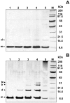
SDS-PAGE analysis of Ub incubated at 37°C in the absence of CuII (A) and with 3 mol equiv of CuII (B) at different incubation time intervals: 2 h (lane 1), 3 d (lane 2), 1 w (lane 3), 2 w (lane 4), 2 mo (lane 5).
Figure 3
Figure 3. Disruption of CuII-stabilized Ub oligomers by addition of chelating agents.
(A) SDS-PAGE of Ub incubated for 2 weeks at 37°C with 3 mol equiv of CuII in 20% TFE (lane 1) and then incubated for 2 h with increasing concentrations of EDTA: 100 µM (lane 2), 200 µM (lane 2), and 300 µM (lane 4); (B) SDS-PAGE of Ub incubated for 2 weeks at 37°C with 3 mol equiv of CuII in 20% TFE (lane 1) and then incubated for 2 h with 300 µM IDA (lane 3). Control experiments performed on Ub incubated in water (lane 2) or in 20% TFE (lane 4) are also reported.
Figure 4
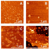
Figure 4. Ub aggregate morphologies for long-term incubations with CuII and/or TFE.
Phase-mode AFM images (A, B, C, D) and TEM micrographs (E, F, G, H) of Ub structures after two months of incubation at 37°C in the absence of CuII (A and E); with 3 mol equiv of CuII (B and F); with 20% TFE (C and G); with 3 mol equiv of CuII in 20% TFE (D and H).
Figure 5
Figure 5. CuII and TFE induced conformational change of Ub.
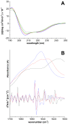
A) Far-UV CD spectra of Ub incubated at 37°C with 3 mol equiv of CuII in 20% TFE for different time intervals: 1 h (blue line); 5 d (red); 1 mo (green); 2 mo (black); B) ATR-FTIR spectrum (absorbance at the top and second derivative at the bottom) in the amide I region of native Ub (blue line) and of Ub incubated with 3 mol equiv of CuII in 20% TFE for 1 w (red) and 2 mo (black).
Figure 6
Figure 6. Hierarchical assembly of Ub aggregates.
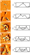
Phase-mode AFM images of Ub structures after incubation at 37°C with 3 mol equiv of CuII in 20% TFE for different time intervals: 1 d (A); 5 d (B); 10 d (C, D); 1 mo (E). (Right panel) Cross-sectional profile, taken along the cyan line, of the corresponding topographic image.
Figure 7
Figure 7. Redox-mediated aggregate disassembly.
A) SDS-PAGE of Ub incubated for 2 weeks at 37°C with 3 mol equiv of CuII in 20% TFE before (lane 1) and after (lane 3) 2 h treatment with ascorbic acid. Control experiments performed on Ub incubated in water (lane 2) or in 20% TFE (lane 4) are also reported; B) Phase-mode AFM image and cross-sectional profile, taken along the cyan line, of Ub incubated for 2 months at 37°C with 3 mol equiv of CuII in 20% TFE after 2 h treatment with ascorbic acid. The inset at the bottom right shows an AFM image and a cross-sectional profile of Ub incubated for 2 weeks at 37°C in 20% TFE.
Figure 8
Figure 8. Paramagnetic CuII broadening effects in NMR spectra of Ub in TFE.
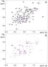
A) Overlay of 1H,15N HSQC spectra of Ub in 50 mM ammonium acetate buffer at pH 6.5 (blue contours) and Ub in the same buffer with 20% TFE (red contours). Backbone amide cross-peaks and side-chains of Gln/Asn are labeled; B) Overlay of 1H,15N HSQC spectra of Ub in 50 mM ammonium acetate buffer at pH 6.5 with 20% TFE before (red contours) and after (blue contours) addition of 0.1 mol equiv of CuII. Cross-peaks that disappear after CuII addition are labeled.
Figure 9
Figure 9. Annular and pore-like assemblies of CuII-stabilized Ub oligomers.
Topographic AFM images of Ub preincubated for two weeks at 37°C with 3 mol equiv of CuII in aqueous solution (A) and then dissolved in 20% TFE (B), in POPC liposomes (C), or in POPC planar bilayers (D).

References

    1. McClellan AJ, Tam S, Kaganovich D, Frydman J. Protein quality control: chaperones culling corrupt conformations. Nat Cell Biol. 2005;7:736–741. - PubMed
    1. Hershko A, Ciechanover A. The ubiquitin system. Annu Rev Biochem. 1998;67:425–479. - PubMed
    1. Pickart CM, Fushman D. Polyubiquitin chains: polymeric protein signals. Curr Opin Chem Biol. 2004;8:610–616. - PubMed
    1. Soto C. Unfolding the role of protein misfolding in neurodegenerative diseases. Nat Rev Neurosci. 2003;4:49–60. - PubMed
    1. Chiti F, Dobson CM. Protein misfolding, functional amyloid, and human disease. Annu Rev Biochem. 2006;75:333–366. - PubMed

Publication types