

High-speed ultrahigh-resolution optical coherence tomography findings in chronic solar retinopathy
- PMID: 19756263
- PMCID: PMC2743270
- DOI: 10.1097/ICB.0b013e3181506993
High-speed ultrahigh-resolution optical coherence tomography findings in chronic solar retinopathy
Abstract
Purpose: To describe ocular findings for a 34-year-old man with chronic solar retinopathy using high-speed ultrahigh-resolution (UHR) optical coherence tomography (OCT).
Methods: Fundus photography, fluorescein angiography, and Stratus OCT (Carl Zeiss Meditec, Inc., Dublin, CA) were performed. A high-speed UHR OCT prototype developed in our ophthalmology clinic was used to obtain detailed images of the retina.
Patients: Two eyes of one patient with chronic solar retinopathy were studied.
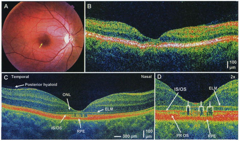
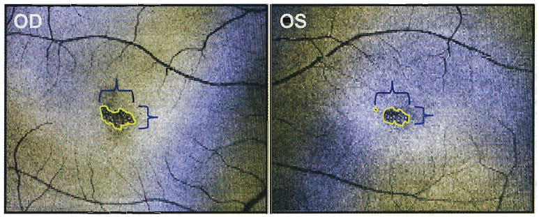
Results: Both Stratus OCT and high-speed UHR OCT demonstrated foveal thinning bilaterally. In addition, high-speed UHR OCT showed distinct hyporeflective disruptions in the photoreceptor inner segment/outer segment junction and photoreceptor outer segments bilaterally. En face OCT images from three-dimensional OCT data sets revealed hyporeflective regions of photoreceptor atrophy in the outer retina.
Conclusions: High-speed UHR OCT showed more detail than standard OCT, and findings were consistent with histopathologic and ultrastructural features that have been reported previously. Solar retinopathy should be studied further with high-speed UHR OCT to determine the short- and long-term effects of solar radiation damage.
Figures
References
-
- Tso MO, La Piana FG. The human fovea after sungazing. Trans Am Acad Ophthalmol Otolaryngol. 1975;79:788–795. - PubMed
-
- Hope-Ross MW, Mahon GJ, Gardiner TA, et al. Ultrastructural findings in solar retinopathy. Eye. 1993;7:29–33. - PubMed
-
- Gass JDM. Stereoscopic Atlas of Macular Diseases: Diagnosis and Treatment. 4th. St. Louis: Mosby; 1997. pp. 760–763.
-
- Garg SJ, Martidis A, Nelson ML, Sivalingam A. Optical coherence tomography of chronic solar retinopathy. Am J Ophthalmol. 2004;137:351–354. - PubMed
Grants and funding
LinkOut - more resources
Full Text Sources
Research Materials
