Clinical Trial
. 2010 Feb;248(2):167-73.
doi: 10.1007/s00417-009-1187-2. Epub 2009 Sep 16.

Venous retinal flow reperfusion mechanisms following radial optic neurotomy with adjunctive intraocular triamcinolone in central retinal vein occlusion

Affiliations
Clinical Trial

Venous retinal flow reperfusion mechanisms following radial optic neurotomy with adjunctive intraocular triamcinolone in central retinal vein occlusion

Pina Fortunato et al. Graefes Arch Clin Exp Ophthalmol. 2010 Feb.

Abstract

Background: To evaluate retinal reperfusion, anastomosis, and compensation following radial optic neurotomy for ischemic nonperfused central retinal vein occlusion.

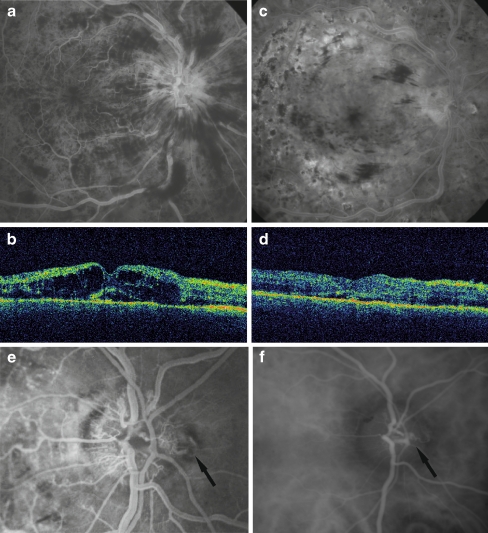
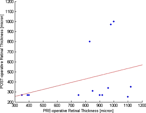
Methods: Prospective, non-comparative, interventional case series of 13 patients with ischemic, nonperfused central retinal vein occlusion who underwent decompression surgery with a radial optic neurotomy and adjunctive intraocular triamcinolone. Patients were followed for 1 year after surgery, and were analyzed in the preoperative and postoperative stages determining: visual acuity by ETDRS chart (LogMar) and retinal thickness with optical coherence tomography. Fluorescein angiography was performed at regular intervals to evaluate the capillary perfusion grade. Intraocular pressure was measured and fundus was examined.

Results: Visual acuity and retinal thickness improved in 10/13 (77%) patients after surgery at 1-year follow-up. Adequate retinal reperfusion was achieved in ten of the 13 eyes. Acute reperfusion occurred in six eyes within 2 weeks of surgery and a shunt vessel at the optic disk developed in four eyes within 4 months. In the remaining three eyes, retinal reperfusion was not observed by fluorescein angiography. No complications were noted in any of the patients.

Conclusion: Surgical decompression promoted mechanical reperfusion of the occluded vessel in 10/13 (77%) cases. In 6/13 patients (46%) reperfusion occurred within 2 weeks of surgery, and in 4/13 patients (31%) collateral vessels formed within 4 months.


Figures

Fig. 1
Fig. 1
Patient 3. a Preoperative fluorescein angiography showing marked edema of the optic disc and venous stasis. b Preoperative foveal thickness measured by optical coherence tomography was 920 µm. Macular edema and serous detachment of the neuroepithelium. Preoperative VA was 0.7 (LogMar). c Postoperative fluorescein angiography 2 months after surgery showing decreased edema and venous stasis. d Postoperatively, foveal thickness decreased to 270 µm, the macular edema disappeared, and the serous detachment was reduced. Postoperative VA was 0.1 (LogMar). e,f Fluorescein angiography and indocyanine green angiography 4 months after surgery showed the anastomosis at the nasal neurotomy site (arrow)
Fig. 2
Fig. 2
Patient 5. a Preoperative fluorescein angiography showing macular edema and venous stasis secondary to central retinal vein occlusion. b Preoperative optical coherence tomography indicates a foveal thickness of 1,100 µm. Preoperative VA was 2.0 (LogMar). c Postoperative fluorescein angiography showing resolution of the venous dilation and tortuosity, and hemorrhage. d Postoperatively, foveal thickness decreased to 250 µm with partial resolution of the edema. Postoperative VA was 0.7 (LogMar). e Fluorescein angiography 4 months after surgery showed the anastomosis at the nasal RON site (arrow)
Fig. 3
Fig. 3
Patient 6. a Preoperative fluorescein angiography showing extensive intraretinal hemorrhages and venous turgidity. b Preoperative foveal thickness measured by optical coherence tomography was 750 µm. Preoperative VA was 0.7 (LogMar). c Postoperative fluorescein angiography showing resolution with normal retinal vasculature. d Postoperative foveal thickness was 270 µm. Postoperative VA was −0.1 (LogMar). e Fundus photograph 4 months after surgery showed anastomosis at the nasal incision site (arrow)
Fig. 4
Fig. 4
Scatter plot of the pre- and post-operative values of Snellen Visual Acuity (the regression line showed is for illustrative purpose only)
Fig. 5
Fig. 5
Scatter plot showing the pre and post-operative values of retinal thickness (µm)

References

    1. The Eye Disease Case-Control Study Group Risk factors for central retinal vein occlusion. Arch Ophthalmol. 1996;114:545–554. - PubMed
    1. Rath EZ, Frank RN, Shin DH, Kim C. Risk factors for retinal vein occlusions: a case-controlled study. Ophthalmology. 1992;99:509–514. - PubMed
    1. Hayreh SS. Retinal vein occlusion. Indian J Ophthalmol. 1994;42:109–132. - PubMed
    1. Hayreh SS. Prevalent misconceptions about acute retinal vascular occlusive disorders. Prog Retin Eye Res. 2005;24:493–519. doi: 10.1016/j.preteyeres.2004.12.001. - DOI - PubMed
    1. Green WR, Chan CC, Hutchins GM, Terry JM. Central retinal vein occlusion: a prospective histopathologic study of 29 eyes in 28 cases. Retina. 2005;25:27–55. doi: 10.1097/00006982-200507001-00008. - DOI - PubMed

Publication types

MeSH terms