

Diagnosing suspected scaphoid fractures: a systematic review and meta-analysis
- PMID: 19756904
- PMCID: PMC2816764
- DOI: 10.1007/s11999-009-1081-6
Diagnosing suspected scaphoid fractures: a systematic review and meta-analysis
Abstract
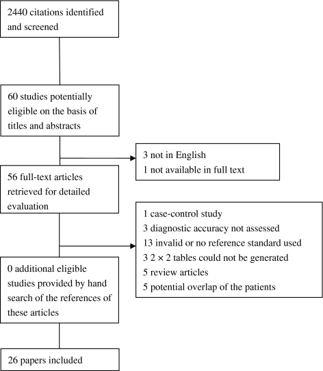
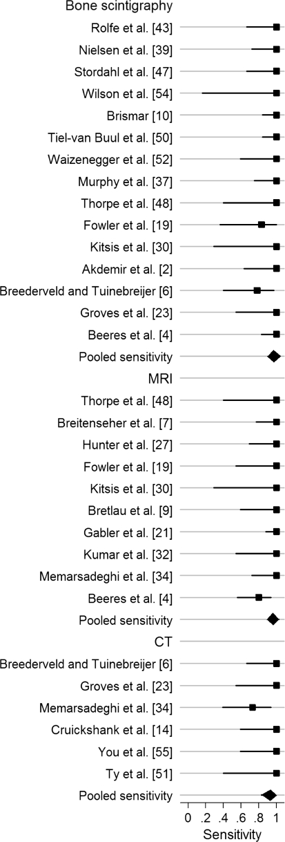
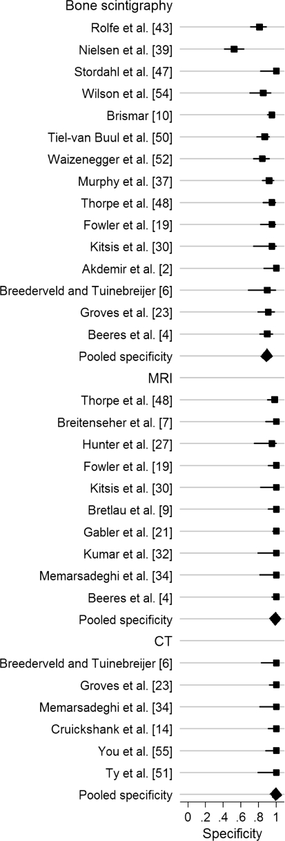
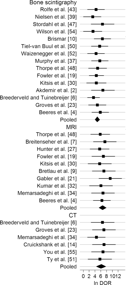
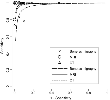
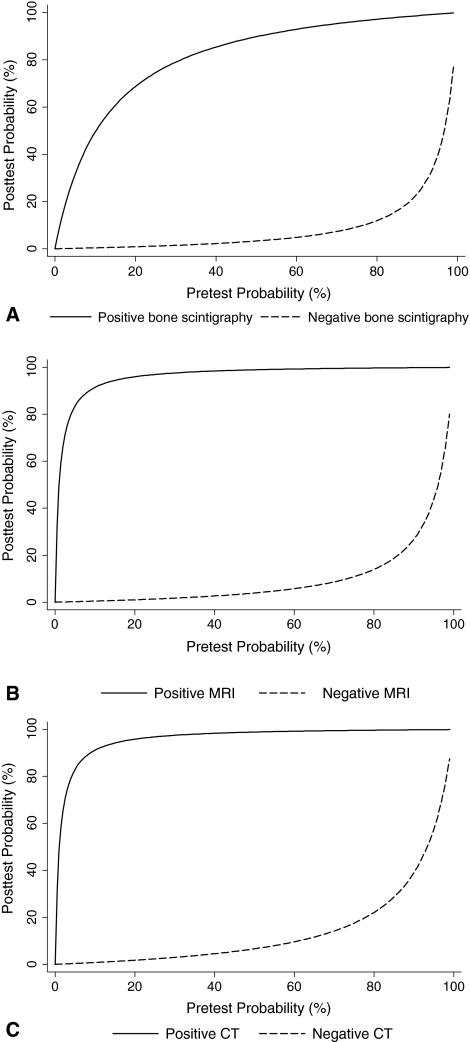
Imaging protocols for suspected scaphoid fractures among investigators and hospitals are markedly inconsistent. We performed a systematic review and meta-analysis to assess and compare the diagnostic performance of bone scintigraphy, MRI, and CT for diagnosing suspected scaphoid fractures. Twenty-six studies were included. Sensitivity, specificity, and diagnostic odds ratio were pooled separately and summary receiver operating characteristic curves were fitted for each modality. Meta-regression analyses were performed to compare these modalities. We obtained likelihood ratios derived from the pooled sensitivity and specificity and, using Bayes' theorem, calculated the posttest probability by application of the tests. The pooled sensitivity, specificity, natural logarithm of the diagnostic odds ratio, and the positive and negative likelihood ratios were, respectively, 97%, 89%, 4.78, 8.82, and 0.03 for bone scintigraphy; 96%, 99%, 6.60, 96, and 0.04 for MRI; and 93%, 99%, 6.11, 93, and 0.07 for CT. Bone scintigraphy and MRI have equally high sensitivity and high diagnostic value for excluding scaphoid fracture; however, MRI is more specific and better for confirming scaphoid fracture. We believe additional studies are needed to assess diagnostic performance of CT, especially paired design studies or randomized controlled trials to compare CT with MRI or bone scintigraphy.
Level of evidence: Level III, diagnostic study. See the Guidelines for Authors for a complete description of levels of evidence.
Figures
Comment in
-
Comparison of imaging techniques for diagnosing suspected scaphoid fractures: a review.Clin J Sport Med. 2010 Sep;20(5):393-4. doi: 10.1097/JSM.0b013e3181f255d9. Clin J Sport Med. 2010. PMID: 20818201 No abstract available.
References
-
- Amrami KK. Radiology corner: diagnosing radiographically occult scaphoid fractures—what’s the best second test? J Am Soc Surg Hand. 2005;5:134–138. doi: 10.1016/j.jassh.2005.05.001. - DOI
-
- Beeres FJ, Rhemrev SJ, Hollander P, Kingma LM, Meylaerts SA, le Cessie S, Bartlema KA, Hamming JF, Hogervorst M. Early magnetic resonance imaging compared with bone scintigraphy in suspected scaphoid fractures. J Bone Joint Surg Br. 2008;90:1205–1209. doi: 10.1302/0301-620X.90B9.20341. - DOI - PubMed
Publication types
MeSH terms
LinkOut - more resources
Full Text Sources
Medical
Research Materials
