. 2009 Dec;11(6):1269-78.
doi: 10.1007/s10544-009-9346-0.

Triangular neuronal networks on microelectrode arrays: an approach to improve the properties of low-density networks for extracellular recording

Affiliations

Triangular neuronal networks on microelectrode arrays: an approach to improve the properties of low-density networks for extracellular recording

Melanie Jungblut et al. Biomed Microdevices. 2009 Dec.

Abstract

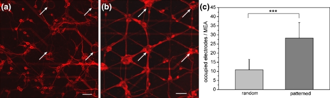
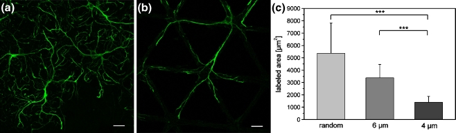
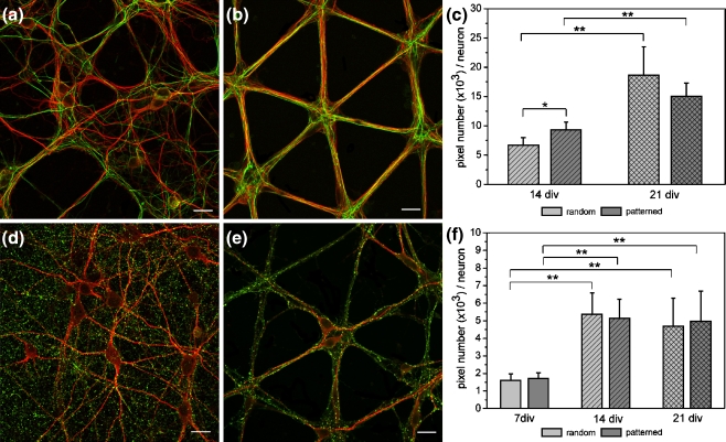
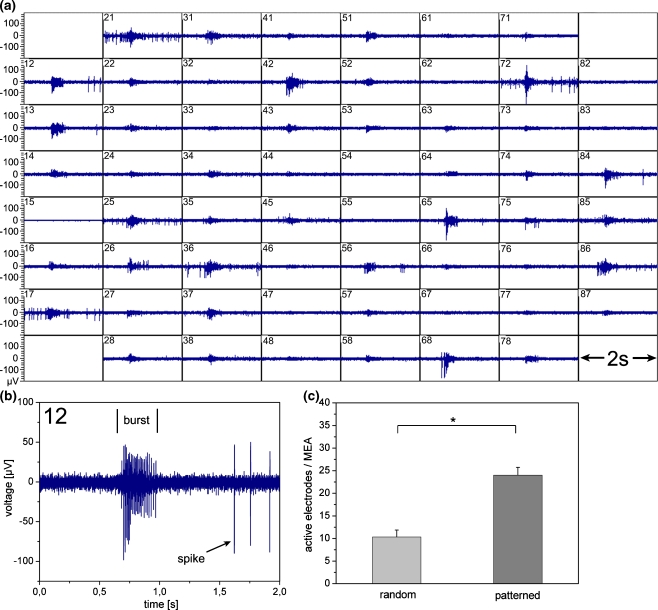
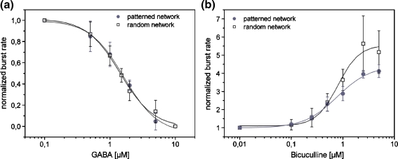
Multi-unit recording from neuronal networks cultured on microelectrode arrays (MEAs) is a widely used approach to achieve basic understanding of network properties, as well as the realization of cell-based biosensors. However, network formation is random under primary culture conditions, and the cellular arrangement often performs an insufficient fit to the electrode positions. This results in the successful recording of only a small fraction of cells. One possible approach to overcome this limitation is to raise the number of cells on the MEA, thereby accepting an increased complexity of the network. In this study, we followed an alternative strategy to increase the portion of neurons located at the electrodes by designing a network in confined geometries. Guided settlement and outgrowth of neurons is accomplished by taking control over the adhesive properties of the MEA surface. Using microcontact printing a triangular two-dimensional pattern of the adhesion promoter poly-D-lysine was applied to the MEA offering a meshwork that at the same time provides adhesion points for cell bodies matching the electrode positions and gives frequent branching points for dendrites and axons. Low density neocortical networks cultivated under this condition displayed similar properties to random networks with respect to the cellular morphology but had a threefold higher electrode coverage. Electrical activity was dominated by periodic burst firing that could pharmacologically be modulated. Geometry of the network and electrical properties of the patterned cultures were reproducible and displayed long-term stability making the combination of surface structuring and multi-site recording a promising tool for biosensor applications.


Figures

Fig. 1
Fig. 1
Neuronal cell cultures on microelectrode arrays (a, b) Labeling of neuronal cell bodies and dendrites using an antibody directed against MAP-2 for random (a) and patterned (b) networks (line width: 6 µm) at 7 div. Some electrodes are highlighted by arrows. Scale bars, 50 µm. (c) Average number of electrodes covered by neuronal cell bodies for random and patterned cell growth at 7 div (n = 12; *** P < 0.001)
Fig. 2
Fig. 2
Astrocyte growth for different culture conditions (a, b) Astrocytes were labeled by antibody staining of GFAP in random (a) and patterned (b) networks at 14 div. Staining of astrocytes in networks with 4 µm wide lines is not shown as almost no staining was present in these cultures. Scale bars, 20 µm. (c) Average labeled area per picture of GFAP-positive structures at 14 div (n = 60; *** P < 0.001)
Fig. 3
Fig. 3
Axonal growth and synaptic density in random and patterned neuronal networks (line width: 6 µm) (a, b) Fluorescence micrographs of neuronal networks showing axonal growth on homogeneously coated (a) and patterned substrate surfaces (b) at 14 div. MAP-2 positive cell bodies and dendrites are depicted in red. Axons were labeled by antibody staining against Neurofilament 160 kD (green). (c) Average pixel number of Neurofilament-positive structures per neuron at 14 and 21 div (n = 30; * P < 0.05; ** P < 0.01). (d, e) Fluorescence micrographs showing synaptic density in cultures after 14 div. Presynaptic sites in random (d) and patterned (e) networks were labeled by immunodetection of Synapsin I (green). Neuronal cell bodies and dendrites were labeled by MAP-2 immunoreactivity (red). (f) Average pixel number per neuron of Synapsin puncta at 7, 14 and 21 div (n = 30; ** P < 0.01). Scale bars, 20 µm
Fig. 4
Fig. 4
Spontaneous electrical activity of mature patterned networks (line width: 6 µm) (a) Electrical activity recorded by the 60 electrodes of a MEA at 24 div. The activity pattern shows single spikes and one burst. Bursts occur synchronized at several electrodes. (b) Neuronal activity detected by electrode 12. Every burst is followed by a silent period of at least 0.5 s before single spikes are observed again. (c) Average number of electrodes detecting neuronal electrical activity for random and patterned networks after 24 div (n = 3; * P < 0.05)
Fig. 5
Fig. 5
GABAergic modulation of the network activity (a) Dose-response curve for GABA-mediated inhibition of spontaneous burst activity. The IC50 was 1.63 µM for patterned networks (line width: 6 µm) and 1.43 µM for random networks of high cell density (n = 3). (b) Increase of spontaneous burst activity in response to the GABAA-receptor antagonist Bicuculline. Bicuculline application resulted in an EC50 of 0.77 µM for patterned networks (line width: 6 µm) and 0.80 µM for random networks of high cell density (n = 4)

References

    1. Angulo CM, Kozlov AS, Charpak S, Audinat E. J. Neurosci. 2004;27:6920. doi: 10.1523/JNEUROSCI.0473-04.2004. - DOI - PMC - PubMed
    1. Boehler MD, Wheeler BC, Brewer GJ. Neuron Glia Biol. 2008;3:127. - PMC - PubMed
    1. Branch DW, Wheeler BC, Brewer GJ, Leckband DE. IEEE Trans. Biomed. Eng. 2000;47:290. doi: 10.1109/10.827289. - DOI - PubMed
    1. Branch DW, Wheeler BC, Brewer GJ. Biomaterials. 2001;22:1035. doi: 10.1016/S0142-9612(00)00343-4. - DOI - PubMed
    1. Brewer GJ, Torricelli JR, Everge EK, Price PJ. J. Neurosc. Res. 1993;35:567. doi: 10.1002/jnr.490350513. - DOI - PubMed

Substances

LinkOut - more resources