Review
. 2009 Nov;16(11):757-68.
doi: 10.1111/j.1365-2893.2009.01194.x. Epub 2009 Sep 15.

GB virus type C interactions with HIV: the role of envelope glycoproteins

Affiliations
Review

GB virus type C interactions with HIV: the role of envelope glycoproteins

Emma L Mohr et al. J Viral Hepat. 2009 Nov.

Abstract

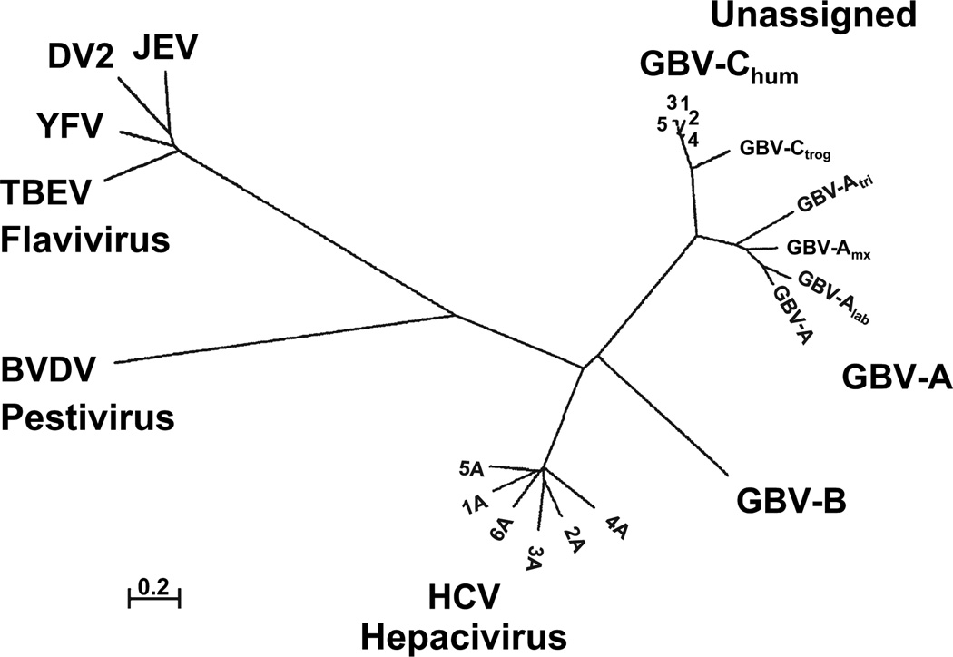
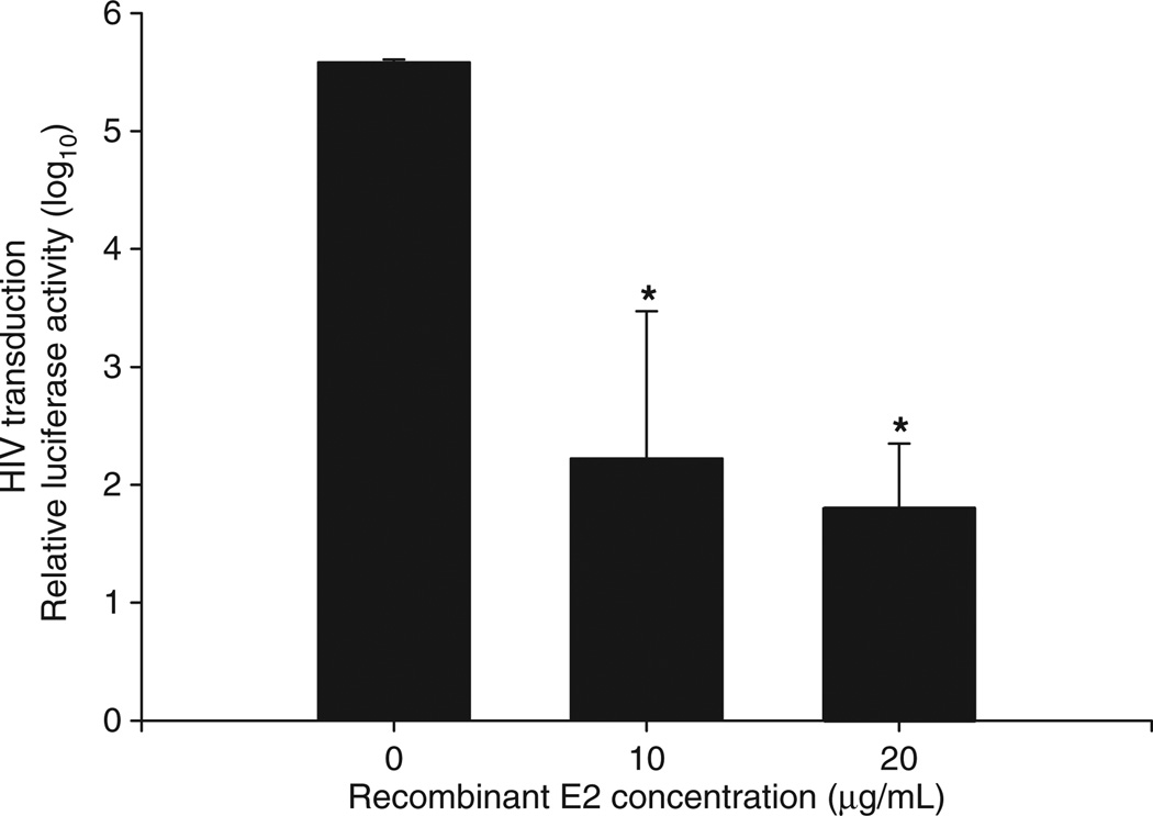
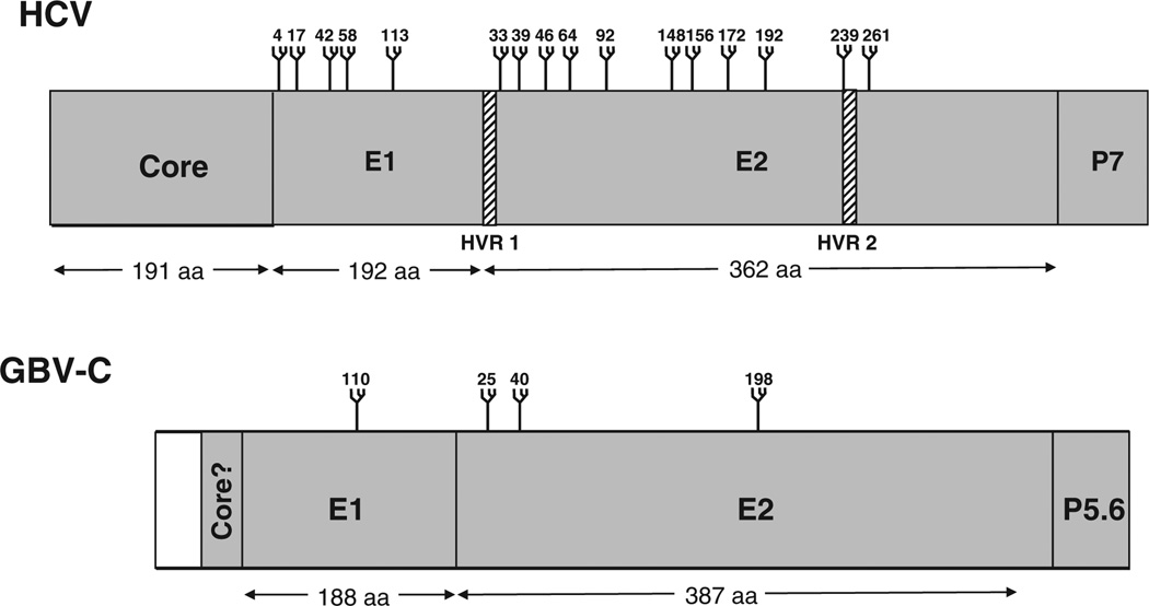
GB virus C/hepatitis G virus (GBV-C/HGV) is the most closely related human virus to hepatitis C virus (HCV). GBV-C is lymphotropic and not associated with any known disease, although it is associated with improved survival in HIV-infected individuals. In peripheral blood mononuclear cells, GBV-C induces the release of soluble ligands for HIV entry receptors (RANTES, MIP-1a, MIP-1b and SDF-1), suggesting that GBV-C may interact with lymphocytes to induce a chemokine and/or cytokine milieu that is inhibitory to HIV infection. Expression of GBV-C envelope glycoprotein E2 in CD4+ T cells or addition of recombinant E2 to CD4 cells recapitulates the HIV inhibition seen with GBV-C infection. Like HCV E2, GBV-C E2 is predicted to be post-translationally processed in the endoplasmic reticulum and is involved with cell binding. The C-termini of GBV-C E1 and E2 proteins contain predicted transmembrane domains sharing features with HCV TM domains. To date, cellular receptor(s) for GBV-C E2 have not been identified. GBV-C E2-mediated HIV inhibition is dose-dependent and HIV replication is blocked at the binding and/or entry step. In addition, a putative GBV-C E2 fusion peptide interferes with HIV gp41 peptide oligomerization required for HIV-1 fusion, further suggesting that GBV-C E2 may inhibit HIV entry. Additional work is needed to identify the GBV-C E2 cellular receptor, characterize GBV-C E2 domains responsible for HIV inhibition, and to examine GBV-C E2-mediated fusion in the context of the entire envelope protein or viral-particles. Understanding the mechanisms of action may identify novel approaches to HIV therapy.


Figures

Fig. 1
Fig. 1
GB virus C prevalence rates in blood donors from various regions of the world. GBV-C viraemia prevalence among 11 391 blood donors was summarized from 50 studies (references available upon request). Viraemia was detected by RT-PCR, and studies only included donors who passed screening procedures with the exception of one Scandinavian study that included donors with normal and high ALT (n = 393 high ALT, n = 184 with normal ALT; the GBV-C prevalence rate was similar in both groups). In developed countries, GBV-C RNA prevalence in blood donors ranged from 0.5 to 5%, compared to 5–18.9% in developing countries. *Caribbean (West Indies).
Fig. 2
Fig. 2
Phylogenetic relationships of the RNA-dependent, RNA polymerase sequences of several members of the family Flaviviridae. Three genera (flavivirus, pestivirus and hepacivirus) and the unassigned GB viruses are shown. Representative isolates from the six hepatitis C virus (HCV), five GBV-C, chimpanzee GBV-C (troglodyte) variant, and four GBV-A genotypes are depicted. The four GBV-A geno-types were identified in Sanguinus nigrocallis (GBV-A), Sanguinus labiatus (GBV-Alab), Aotus Trivirgatus (GBV-Alab), and a ‘callithrix hybrid’ (jacchus-penicillata cross) (GBV-Amyx). BVDV, bovine viral diarrhoea virus; BVDV; YFV, yellow fever virus (17D vaccine strain); TBEV, tick-borne encephalitis virus; DV, dengue virus (serotype 2) and JEV, Japanese encephalitis virus. 0.2, distance representing 0.2 amino acids substitutions per position.
Fig. 3
Fig. 3
Genome organization and proteolytic processing of hepatitis C virus (HCV) and GB virus C (GBV-C). HCV and GBV-C both contain 5′ nontranslated regions (NTR) containing internal ribosomal entry sites directing translation of polyproteins. The polyproteins are post-translationally processed into structural proteins [core (C), envelope glycoproteins (E1 and E2)] and an ion channel P7 (P5.6 for GBV-C) by cellular signal peptidases. Nonstructural proteins (NS) 2 and 3 are cleaved by NS2 protease, while the remaining cleavage sites are processed by the serine protease domain within NS3, in conjunction with the NS4A cofactor.
Fig. 4
Fig. 4
GB virus C envelope glycoprotein 2 (E2) inhibits early steps in the HIV life cycle. Ghost cells expressing CD4, CCR5 and CXCR4 were incubated with recombinant E2 at 4 °C for 4 h prior to transduction with HIV pseudotyped particles bearing a luciferase reporter. GBV-C E2 inhibited transduction of HIV pseudotyped particles in a dose–dependent fashion (*P < 0.01; t-test).
Fig. 5
Fig. 5
Comparison of hepatitis C virus (HCV) and GB virus C (GBV-C) structural proteins and glycosylation sites. The relative size and predicted N-linked glycosylation sites on HCV and GBV-C core and envelope glycoproteins (E1 and E2) proteins are shown (http://www.cbs.dtu.dk/services/NetNGlyc/). The GBV-C core protein is depicted as residing at the N-terminus of the polyprotein. Grey represents the GBV-C Core if the AUG at position 462 serves as initiation codon, while blue represents initiation at the AUG at 555.
Fig. 6
Fig. 6
Functional mapping of the GB virus C envelope glycoprotein E2. The predicted GBV-C E2 transmembrane domain (TM) resides between amino acids 343 and 371 (a). Two putative peptide regions within E2 contain membrane interacting or fusion functions (347–364, within the TM) and 267–298. The M6 monoclonal antibody blocks E2 binding to cells, and recognizes a linear epitope including amino acids 276–292 within the putative fusion peptide (a). HCV hydrophobic TM regions on both E1 and E2 appear to be too short to serve as classical transmembrane α-helices, and are predicted to fold as two antiparallel β-strands in a hairpin structure (b). The amphiphilic α-helix upstream of the hairpin is predicted to stabilize the hairpin on the ER membrane (b). Hydrophobic cluster analysis of HCV, human and chimpanzee variants of GBV-C (GBVChum and GBV-Ctrog respectively) found that the E1 C-termini of these three viruses are similar (c) and share the putative structure shown in panel B. However, only the HCV E2 protein had this type of C terminus, while GBV-C contained helix breaking proline residues upstream from the TM region (c).

References

    1. Simons JN, Leary TP, Dawson GJ, et al. Isolation of novel virus-like sequences associated with human hepatitis. Nat Med. 1995;1:564–569. - PubMed
    1. Linnen J, Wages J, Zhang-Keck Z-Y, et al. Molecular cloning and disease association of hepatitis G virus: a transfusion-transmissible agent. Science. 1996;271:505–508. - PubMed
    1. Deka N, Sharma MD, Mukerjee R. Isolation of the novel agent from human stool samples that is associated with sporadic non-A, non-B hepatitis. J Virol. 1994;68:7810–7815. - PMC - PubMed
    1. Schaluder GG, Dawson GJ, Simons JN, et al. Molecular and serologic analysis in the transmission of the GB hepatitis agents. J Med Virol. 1995;46:81–90. - PubMed
    1. Alter HJ. G-pers creepers, where’d you get those papers? A reassessment of the literature on the hepatitis G virus. Transfusion. 1997;37:569–572. - PubMed

Publication types

MeSH terms

Substances