. 2009 Dec;90(6):605-14.
doi: 10.1111/j.1365-2613.2009.00678.x. Epub 2009 Sep 15.

PDEs1-5 activity and expression in tissues of cirrhotic rats reveal a role for aortic PDE3 in NO desensitization

Affiliations

PDEs1-5 activity and expression in tissues of cirrhotic rats reveal a role for aortic PDE3 in NO desensitization

Rima Tahseldar-Roumieh et al. Int J Exp Pathol. 2009 Dec.

Abstract

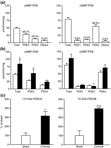
Liver cirrhosis is associated with increased nitric oxide (NO) production in the vasculature. We have previously demonstrated that aorta from rats with liver cirrhosis have a reduced relaxant response to NO donors that is corrected by DMPPO, a PDE5-specific inhibitor. Vasodilator responses to DMPPO itself were also reduced in rings from cirrhotic rats. These results supported previous suggestions that upregulation of PDE5 in liver cirrhosis might contribute to renal sodium retention, and consequently modulate vascular reactivity in the context of increased NO production (Tahseldar-Roumieh et al. in Am. J. Physiol. Heart Circ. Physiol. 290, H481-H488, 2006). Here, we investigated the possible alteration in activity and expression of cyclic nucleotide phosphodiesterase PDE1-PDE5 in kidney and vascular tissues in rats 4 weeks after bile duct ligation. The kidney of rats with cirrhosis had increased activity of PDE1 and PDE4 but not PDE5, and increased expression of PDE1A. Unexpectedly and interestingly, there was no change in cirrhotic aorta PDE5, but an increase in PDE3 and PDE4 activity associated with increased expression of PDE3A and PDE3B. Cilostamide, a specific PDE3 inhibitor, corrected the decreased response to an NO donor in isolated aorta from cirrhotic rats, suggesting that the difference in response to NO donors was due to differences in PDE3-induced hydrolysis of cGMP or to cGMP-induced inhibition of PDE3, rather than to differences in PDE5 contribution. In conclusion, these changes in PDE isozymes could greatly contribute to NO desensitization and to the regulation of vascular and renal function in liver cirrhosis.


Figures

Figure 1
Figure 1
Kidney PDE activity pattern and protein expression of sham-operated (SO) and cirrhotic rats. (a) PDE activity pattern in kidney of SO rat. cAMP–PDE and cGMP–PDE specific activities were assessed as described in Methods. Results are expressed as the mean ± SEM of four independent experiments. The proportion of the different isozymes is expressed as % of total activity. (b) Effect of cirrhosis on PDE activity and isozyme contribution in kidney. cAMP–PDE and cGMP–PDE specific activities were assessed in SO (□) and cirrhotic (▪) rats. Results are expressed as the mean ± SEM of four independent experiments. *P < 0.05; **P < 0.01 versus SO. (c) Effect of cirrhosis on PDE1 expression in kidney. PDE1A expression in kidney is shown for SO (□) and cirrhotic (▪) rats. Histograms showing densitometric analysis are expressed as the mean ± SEM of four independent experiments. ***P < 0.001 versus SO.
Figure 2
Figure 2
Renal cortex PDE activity pattern and protein expression of sham-operated (SO) and cirrhotic rats. (a) PDE activity pattern in renal cortex of SO rat. cAMP–PDE and cGMP–PDE specific activities were assessed as described in Methods. Results are expressed as the mean ± SEM of four independent experiments. The proportion of the different isozymes is expressed as % of total activity. (b) Effect of cirrhosis on PDE activity and isozyme contribution in renal cortex. cAMP–PDE and cGMP–PDE specific activities were assessed in SO (□) and cirrhotic (▪) rats. Results are expressed as the mean ± SEM of four independent experiments. *P < 0.05 versus SO. (c) Effect of cirrhosis on PDE3 expression in renal cortex. PDE3A and PDE3B expressions in renal cortex are shown for SO (□) and cirrhotic (▪) rats. Histograms showing densitometric analysis are expressed as the mean ± SEM of four independent experiments. ***P < 0.001 versus SO.
Figure 3
Figure 3
Mesenteric arteries PDE activity pattern and protein expression of sham-operated (SO) and cirrhotic rats. (a) PDE activity pattern in mesenteric arteries of SO rat. cAMP–PDE and cGMP–PDE specific activities were assessed as described in Methods. Results are expressed as the mean ± SEM of four independent experiments. The proportion of the different isozymes is expressed as % of total activity. (b) Effect of cirrhosis on PDE activity and isozyme distribution in mesenteric arteries. cAMP–PDE and cGMP–PDE specific activities were assessed in SO (□) and cirrhotic (▪) rats. Results are expressed as the mean ± SEM of four independent experiments. *P < 0.05; **P < 0.01 versus SO. (c) Effect of cirrhosis on PDE3 and PDE4 expression in mesenteric arteries. PDE3B and PDE4D expressions in mesenteric arteries are shown for SO (□) and cirrhotic (▪) rats. Histograms showing densitometric analysis are expressed as the mean ± SEM of four independent experiments. **P < 0.01; ***P < 0.001 versus SO.
Figure 4
Figure 4
Aorta PDE activity pattern and protein expression of sham-operated (SO) and cirrhotic rats. (a) PDE activity pattern in aorta of SO rat. cAMP–PDE and cGMP–PDE specific activities were assessed as described in Methods. Results are expressed as the mean ± SEM of four independent experiments. The proportion of the different isozymes is expressed as % of total activity. (b) Effect of cirrhosis on PDE activity and isozyme contribution in aorta. cAMP–PDE and cGMP–PDE specific activities were assessed in SO (□) and cirrhotic (▪) rats. Results are expressed as the mean ± SEM of four independent experiments. *P < 0.05; **P < 0.01 versus SO. (c) Effect of cirrhosis on PDE3 expression in aorta. PDE3A and PDE3B expressions in aorta are shown for SO (□) and cirrhotic (▪) rats. Histograms showing densitometric analysis are expressed as the mean ± SEM of four independent experiments. *P < 0.05; ***P < 0.001 versus SO.
Figure 5
Figure 5
Vasorelaxation studies on endothelium-denuded aortic rings from sham-operated (SO) and cirrhotic rats. (a) Cumulative concentration response curves (CCRC) for cilostamide on aortic rings from SO (□, dotted line; n = 9) and cirrhotic (▪, solid line; n = 10,) rats. (b) CCRC for nitroglycerin (NG) in aortic rings from SO and cirrhotic rats in absence of cilostamide. Sham-operated (□, dotted line; n = 30) and cirrhotic (▪, solid line; n = 33) aortic rings. (c) CCRC for NG in aortic rings from SO (□, dotted line; n = 14) and cirrhotic (▪, solid line; n = 18) rats in presence of cilostamide (0.1 μM), a PDE3-specific inhibitor. Endothelium-denuded aortic rings were precontracted with 1 μM phenylephrine as indicated in Methods. Values are means % of PE-induced tension ±SEM of n experiments. (a) *P < 0.05 by t-test for BDL vs. SO. (b) P < 0.0001 by two-way anova, comparing the overall response in the two groups and ***P < 0.001 for BDL vs. SO.

References

    1. Ahloulay M, Bankir L, Lugnier C, et al. Cyclic AMP-phosphodiesterases inhibitor improves sodium excretion in rats with cirrhosis and ascites. Liver Int. 2005;25:403–409. - PubMed
    1. Angeli P, Jimenez W, Veggian R, et al. Increased activity of guanosine 3′-5′-cyclic monophosphate phosphodiesterase in the renal tissue of cirrhotic rats with ascites. Hepatology. 2000;31:304–310. - PubMed
    1. Atucha NM, Nadal FJ, Iyu D, et al. Role of vascular nitric oxide in experimental liver cirrhosis. Curr. Vasc. Pharmacol. 2005;3:81–85. - PubMed
    1. Bernardi M, Di Marco C, Trevisani F, et al. Renal sodium retention during upright posture in preascitic cirrhosis. Gastroenterology. 1993;105:188–193. - PubMed
    1. Blendis L, Wong F. The hyperdynamic circulation in cirrhosis: an overview. Pharmacol. Ther. 2001;89:221–231. - PubMed