. 2009 Nov;13(11):488-95.
doi: 10.1016/j.tics.2009.08.005. Epub 2009 Sep 14.

Topographic maps in human frontal and parietal cortex

Affiliations

Topographic maps in human frontal and parietal cortex

Michael A Silver et al. Trends Cogn Sci. 2009 Nov.

Abstract

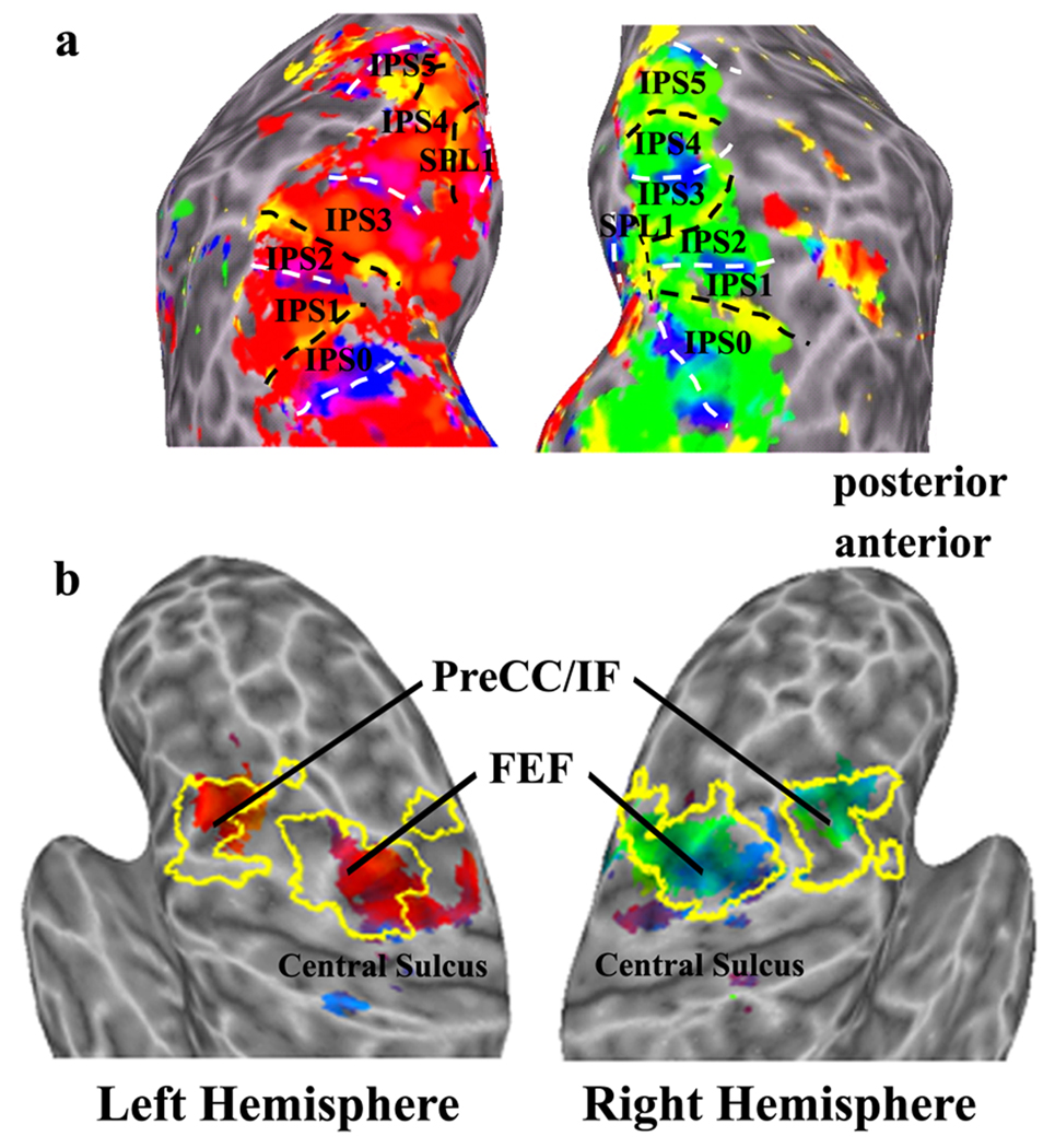
Retinotopic mapping of functional magnetic resonance (fMRI) responses evoked by visual stimuli has resulted in the identification of many areas in human visual cortex and a description of the organization of the visual field representation in each of these areas. These methods have recently been employed in conjunction with tasks that involve higher-order cognitive processes such as spatial attention, working memory, and planning and execution of saccadic eye movements. This approach has led to the discovery of multiple areas in human parietal and frontal areas, each containing a topographic map of visual space. In this review, we summarize the anatomical locations, visual field organization, and functional specialization of these new parietal and frontal topographic cortical areas. The study of higher-order topographic cortex promises to yield unprecedented insights into the neural mechanisms of cognitive processes and, in conjunction with parallel studies in non-human primates, into the evolution of cognition.


Figures

Figure 1
Figure 1. Topographic maps in ventral visual cortex (A) and the lateral geniculate nucleus (B)
A: Flattened surface reconstructions of early and ventral visual cortical areas in the right hemisphere of a single subject. The left panel shows the polar angle maps; the right panel shows the eccentricity maps. White lines denote area boundaries, which are formed by phase angles at or close to the upper (dotted) or lower (dashed) vertical meridian. Pink lines denote the reversal in eccentricity between V4 and VO-1. Asterisks indicate foveal representations. Adapted with modification from Ref [15]. B: Polar angle and eccentricity maps in the LGN are shown for both hemispheres of a single subject. The central panel shows an anatomical image in the coronal plane through the posterior thalamus. The black boxes indicate the anatomical locations of the panels to the left and right. Details of the polar angle maps in the left (L) and right (R) LGNs are shown in the near left and right columns, arranged in several consecutive slices from anterior (A) to posterior (P). The eccentricity maps are shown in the far left and right columns and are spatially coregistered with the polar angle maps. Adapted from Ref [16], in modified form. The color code indicates the phase of the fMRI response and labels the portion of the visual field to which the voxel is most responsive, as depicted in the visual field color legends in panels A and B.
Figure 2
Figure 2. Topographic areas in human parietal (A) and frontal (B) cortex
Activations from a single subject overlaid on inflated left and right cortical hemispheres. The color code is shown only for those voxels whose responses were correlated with the fundamental frequency of saccade direction in a memory-guided saccade task (p <0.001). The responses are lateralized such that the right visual field is represented in the left hemisphere, whereas the left visual field is represented in the right hemisphere. In parietal cortex (A), area boundaries correspond to the alternating representation of either the upper or lower vertical meridian. In frontal cortex (B), the extent of the activations evoked by saccadic eye movements is outlined in yellow, indicating that the topographic areas fall within the regions of the superior and inferior frontal eye fields. Adapted with modifications from Refs [21,49].

References

    1. Kaas JH. Topographic maps are fundamental to sensory processing. Brain Res. Bull. 1997;44:107–112. - PubMed
    1. Inouye T. Die Sehstörungen bei Schussverletzungen der kortikalen Sehsphäre nach Beobachtungen an Versundeten der letzten Japanische Kriege. Wilhelm Engelmann; 1909.
    1. Glickstein M, Fahle M. Visual Disturbances Following Gunshot Wounds of the Cortical Visual Area. Oxford University Press; 2000.
    1. Engel SA, et al. fMRI of human visual cortex. Nature. 1994;369:525. - PubMed
    1. Wandell BA, et al. Visual field maps in human cortex. Neuron. 2007;56:366–383. - PubMed

Publication types