Review
. 2010 Mar-Apr;16(2):205-24.
doi: 10.1093/humupd/dmp032. Epub 2009 Sep 15.

Phenotyping male infertility in the mouse: how to get the most out of a 'non-performer'

Affiliations
Review

Phenotyping male infertility in the mouse: how to get the most out of a 'non-performer'

Claire L Borg et al. Hum Reprod Update. 2010 Mar-Apr.

Abstract

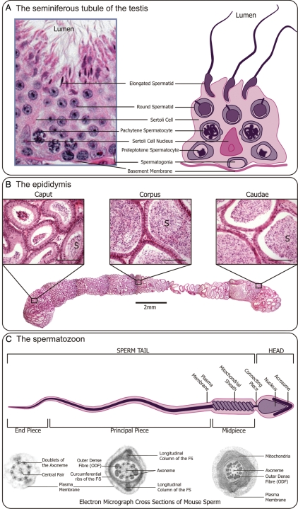
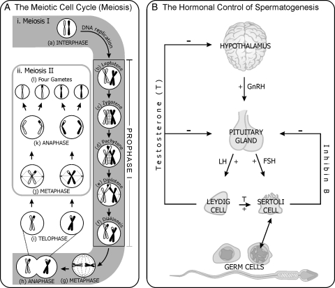
Background: Functional male gametes are produced through complex processes that take place within the testis, epididymis and female reproductive tract. A breakdown at any of these phases can result in male infertility. The production of mutant mouse models often yields an unexpected male infertility phenotype. It is with this in mind that the current review has been written. The review aims to act as a guide to the 'non-reproductive biologist' to facilitate a systematic analysis of sterile or subfertile mice and to assist in extracting the maximum amount of information from each model.

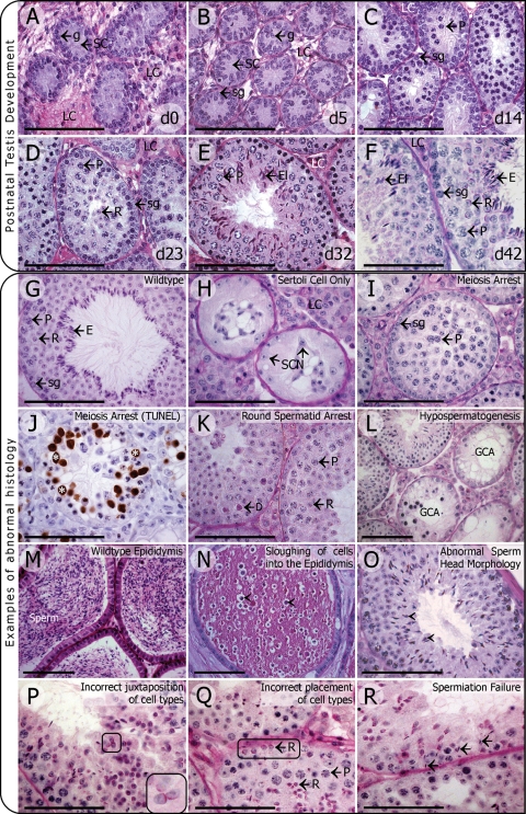
Methods: This is a review of the original literature on defects in the processes that take a mouse spermatogonial stem cell through to a fully functional spermatozoon, which result in male infertility. Based on literature searches and personal experience, we have outlined a step-by-step strategy for the analysis of an infertile male mouse line.

Results: A wide range of methods can be used to define the phenotype of an infertile male mouse. These methods range from histological methods such as electron microscopy and immunohistochemistry, to hormone analyses and methods to assess sperm maturation status and functional competence.

Conclusion: With the increased rate of genetically modified mouse production, the generation of mouse models with unexpected male infertility is increasing. This manuscript will help to ensure that the maximum amount of information is obtained from each mouse model and, by extension, will facilitate the knowledge of both normal fertility processes and the causes of human infertility.


Figures

Figure 1:
Figure 1:
(A) The seminiferous tubule of the testis (a cross-section): the Sertoli cells provide support and nutrients for the developing germ cells. Sertoli cells also form the blood–testis barrier between adjacent Sertoli cells, which functionally divide the seminiferous epithelium into the basal and luminal compartments. Spermatogonia, the self-renewing stem cells of the testis, are associated with the basement membrane of the tubule. As the germ cells develop, from spermatocytes to spermatids, they move progressively closer to the lumen of the tubule where they are released in a process known as spermiation. (B) The epididymis: periodic Acid Schiff (PAS) stained epididymal sections, scale bar = 100 µm. Sperm released from the rete testis enter the efferent ducts, travel through the caput, corpus and caudal epididymal regions, during which time they undergo epididymal maturation. The epididymis ends at the vas deferens (not shown). S=spermatozoa. (C) The Spermatozoon: the spermatozoon is made up of two main regions, the head and the tail. The anterior portion of the head is covered by the acrosomal cap and the head is joined to the tail by the connecting piece. The tail is divided into three regions: the midpiece; principal piece; and the end-piece. The electron micrographs showing cross-sections (not to scale) of each region highlights the main components of the tail structure: the axoneme; outer dense fibers (ODF); and the mitochondrial sheath (midpiece) and fibrous sheath (FS) (principal piece). The end-piece consists solely of the axoneme and plasma membrane.
Figure 2:
Figure 2:
(A) The meiotic cell cycle: chromosomes replicate during interphase (a) of meiosis I (i). Following interphase, the chromosomes move into prophase I, beginning with the first phase, leptotene (b), where the chromosomes remain unpaired, but search each other out. The synaptonemal complex forms during zygotene (c) and homologous chromosomes begin to pair and the chromosomes become compact. Crossing-over occurs during pachytene (d) and the chromosomes are held together by sites of recombination well into diplotene (e). The chromosomes are completely separated during diakinesis (f) where they are pulled to separate poles of the cell. During metaphase (g) of meiosis I, chromosomes are pulled to separate poles by the spindle fibers. The cell divides at the end of anaphase (h), and contains intact/joined sister chromatids. Two separate cells are present at telophase (i). During metaphase (j) of meiosis II (ii), the sister chromatids are pulled to opposite poles of the cell and they separate during anaphase (k). At the end of meiosis II, four haploid gametes (l) have formed from each leptotene spermatocyte. (B) The hormonal control of spermatogenesis: a schematic representation of the hypothalamic–pituitary–testis axis in adulthood. Hypothalamic gonadotrophin-releasing hormone (GnRH) stimulates the pituitary to secrete the follicle stimulating hormone (FSH) and the lutenizing hormone (LH). LH stimulates Leydig cells to produce testosterone (T). FSH and testosterone directly stimulate the Sertoli cells activity, which in turn regulates germ cell development. GnRH is under negative feedback control by testosterone. Pituitary secretion of LH and FSH is under feedback control by testosterone. Inhibin B is produced by the Sertoli cells and causes selective inhibition of FSH production.
Figure 3:
Figure 3:
(A–F) Post-natal testis development: PAS stained sections of mouse testis at varying post-natal days, showing the most mature cell types present. Scale bar = 100 µm. (A) Day 0. Centrally located gonocytes (g) surrounded by Sertoli cells (SC). (B) Day 5. Spermatogonia (sg) at the basement membrane surrounded by Sertoli cells. The occasional gonocyte may remain towards the centre of the tubule. (C) Day 14. Spermatogonia at the basement membrane and the beginning of meiosis with the primary spermatocytes (pachytene) (P) surrounded by Sertoli cells. (D) Day 23. Cells exiting meiosis are termed round spermatids (R), which begin to undergo the process of spermiogenesis towards the centre of the tubule. (E) Day 32. Elongating spermatids (El), undergoing the striking morphological changes to become a fully functional spermatozoon, line the centre of the tubule, anchored by the Sertoli cells. (F) Day 42 (Adult). All cell types are present in the testis forming defined cellular associations between sub-types of spermatogonia, meiotic and post-meiotic cells, called ‘stages’. This tubule at the right contains spermatogonia, pachytene spermatocytes, roundspermatids and elongated spermatids (E) prior to spermiation (release from the testis). G-R. Examples of abnormal testis phenotypes: PAS stained sections of mouse testes showing abnormal phenotypes. All testes were taken from adult mice. Scale bar = 100 µm. (G) A Wild-type testis, showing all cell types in correct numbers. (H) A Sertoli cell only (SCO) testis, no germ cells are present, and only Sertoli cells and their nuclei (SCN) are observed. (I) A meiosis arrest testis, showing an arrest at the pachytene stage of meiosis I (P). (J) TUNEL staining of a meiosis arrest testis, showing an abundance of dying (apoptotic) cells stained in brown (*). (K) A round spermatid arrest testis, no cells past the round spermatid stage (R) are present. Dying cells can be seen (D).(L) A hypospermatogenic testis, wherein there are normal tubule cross-sections alongside abnormal cross-sections, in this case showing germ cell arrest (GCA). (M) An adult wild-type epididymis, displaying an abundance of sperm (S) in the caudal epididymal region. (N) Sloughing of testicular cells into the epididymis, no mature sperm can be seen in the caudal epididymal region, however, a large number of round cells that have prematurely left the testis are observed (arrowheads). (O) A testis containing elongating spermatids with abnormal sperm head morphology, sperm that are abnormally shaped (arrowheads) when compared with the classical falciform (hook) shaped sperm heads seen in the wild-type testis (G). (P) seminiferous epithelium containing incorrectly juxtaposed cell types, disorientation of round spermatids (circled, inset). The pink stained developing acrosomes should be facing the same direction. (Q) Seminiferous epithelium containing an incorrect placement of cell types, round spermatids are seen adjacent to the basement membrane (circled), whereas they should be seen approximately halfway towards the lumen. (R) Seminiferous epithelium containing retained elongated spermatids (arrows) are being drawn to the basement membrane (spermiation failure).

References

    1. Abel MH, Wootton AN, Wilkins V, Huhtaniemi I, Knight PG, Charlton HM. The effect of a null mutation in the follicle-stimulating hormone receptor gene on mouse reproduction. Endocrinology. 2000;141:1795–1803. - PubMed
    1. Abel MH, Baker PJ, Charlton HM, Monteiro A, Verhoeven G, De Gendt K, Guillou F, O'Shaughnessy PJ. Spermatogenesis and Sertoli cell activity in mice lacking Sertoli cell receptors for follicle-stimulating hormone and androgen. Endocrinology. 2008;149:3279–3285. - PMC - PubMed
    1. Abou-Haila A, Fain-Maurel MA. Regional differences of the proximal part of mouse epididymis: morphological and histochemical characterization. Anat Rec. 1984;209:197–208. - PubMed
    1. Abou-Haila A, Tulsiani DR. Mammalian sperm acrosome: formation, contents, and function. Arch Biochem Biophys. 2000;379:173–182. - PubMed
    1. Aitken RJ, Paterson M, Fisher H, Buckingham DW, van Duin M. Redox regulation of tyrosine phosphorylation in human spermatozoa and its role in the control of human sperm function. J Cell Sci. 1995;108:2017–2025. - PubMed

Publication types