. 2009 Nov 20;284(47):32895-905.
doi: 10.1074/jbc.M109.000208. Epub 2009 Sep 15.

Isolation and characterization of patient-derived, toxic, high mass amyloid beta-protein (Abeta) assembly from Alzheimer disease brains

Affiliations

Isolation and characterization of patient-derived, toxic, high mass amyloid beta-protein (Abeta) assembly from Alzheimer disease brains

Akihiko Noguchi et al. J Biol Chem. .

Abstract

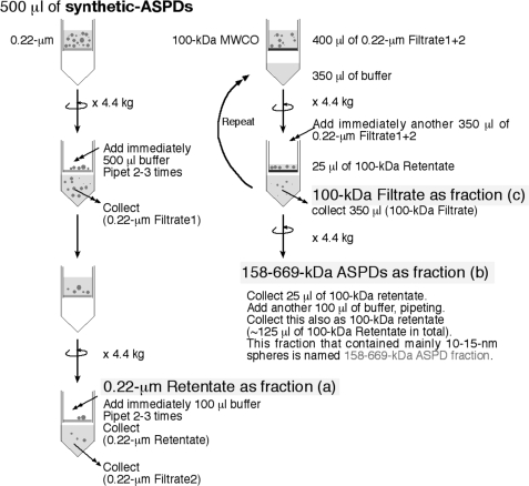
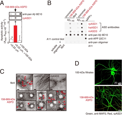
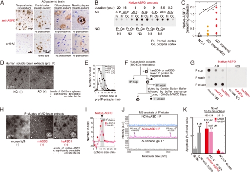
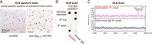
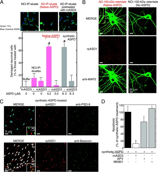
Amyloid beta-protein (Abeta) assemblies are thought to play primary roles in Alzheimer disease (AD). They are considered to acquire surface tertiary structures, not present in physiologic monomers, that are responsible for exerting toxicity, probably through abnormal interactions with their target(s). Therefore, Abeta assemblies having distinct surface tertiary structures should cause neurotoxicity through distinct mechanisms. Aiming to clarify the molecular basis of neuronal loss, which is a central phenotype in neurodegenerative diseases such as AD, we report here the selective immunoisolation of neurotoxic 10-15-nm spherical Abeta assemblies termed native amylospheroids (native ASPDs) from AD and dementia with Lewy bodies brains, using ASPD tertiary structure-dependent antibodies. In AD patients, the amount of native ASPDs was correlated with the pathologic severity of disease. Native ASPDs are anti-pan oligomer A11 antibody-negative, high mass (>100 kDa) assemblies that induce degeneration particularly of mature neurons, including those of human origin, in vitro. Importantly, their immunospecificity strongly suggests that native ASPDs have a distinct surface tertiary structure from other reported assemblies such as dimers, Abeta-derived diffusible ligands, and A11-positive assemblies. Only ASPD tertiary structure-dependent antibodies could block ASPD-induced neurodegeneration. ASPDs bind presynaptic target(s) on mature neurons and have a mode of toxicity different from those of other assemblies, which have been reported to exert their toxicity through binding postsynaptic targets and probably perturbing glutamatergic synaptic transmission. Thus, our findings indicate that native ASPDs with a distinct toxic surface induce neuronal loss through a different mechanism from other Abeta assemblies.


Figures

SCHEME 1.
SCHEME 1.
Fractionation of the most toxic 158–669-kDa ASPDs by two-step filtrations.
FIGURE 1.
FIGURE 1.
Characterization of ASD antibodies. A, evaluation of two-step filtered fractions (0.22-μm retentates, the 158–669-kDa ASPDs, and 100-kDa filtrates; see Scheme 1 under “Experimental Procedures”) by dot blotting using rpASD1 and 6E10 (upper panel) and by toxicity assays using rat primary septal cultures (lower panel; mean ± S.D.; Games-Howell post hoc test, *, p < 0.001, n = 6). B, dot blotting of Aβ and APP (5 ng/dot). Synthetic ASPDs were prepared in vitro either from Aβ-(1–40) or Aβ-(1–42) as described (7). Purified 158–669-kDa ASPD fraction was recovered in 100-kDa retentates as in A. Unlike anti-APP-(66–81) (22C11), anti-Aβ-(1–16) (6E10), or A11 antibody, ASD antibodies selectively detected synthetic ASPDs and the 158–669-kDa ASPDs. The control blot membrane for A11 was provided by Invitrogen (supplemental Experimental Procedures). C, immuno-TEM analysis. Arrows show the secondary antibody-conjugated immunogold. 6E10 detected the 158–669-kDa ASPDs weakly, probably because of its low affinity for synthetic ASPDs. rpASD1 and mASD3 showed little reactivity to fibrils but clearly detected the 158–669-kDa ASPDs. Bar, 20 nm. D, rpASD1 detected intense signals in 27-DIV mature rat hippocampal neurons treated with the 158–669-kDa ASPDs (in A) for 30 min but did not detect signals in those treated with the 100-kDa filtrates containing monomers and Aβ-(1–42) assemblies with mass <100 kDa. Z-stack images are shown (supplemental Experimental Procedures).
FIGURE 2.
FIGURE 2.
Isolation of native ASPDs. A, AD brains were stained with rpASD1 (5 μg/ml) or anti-Aβ1–42 C-terminal antibody (0.5 μg/ml; 2 μg/ml for cryosections). B and C, dot blotting of 100-kDa retentates (>100 kDa) of AD or NCI brain extracts (1 μg of soluble extracts/dot) using rpASD1 (Scheffé post hoc test; **, p = 0.0011; *, p = 0.0388). Fr, frontal cortex; Oc, occipital cortex. D and E, TEM images (D) and particle analysis of 100-kDa retentates (n = 3; 10 randomly selected fields) (E). F and G, method for immunoprecipitation (IP) (F) and dot blotting (using rpASD1) of IP supernatants (sup), wash, and eluate fractions. IPs were performed using haASD1, mASD3, or mouse IgG (G). H and I, TEM images (inset, bar, 10 nm) (H) and particle analysis of IP eluates (n = 3; 15 randomly selected fields, background (a small amount of spheres <10 nm contained in eluate with buffers)-subtracted data are shown) (I). J, representative MALDI-TOF/MS data. Aβ-(1–40) and Aβ-(1–42) were detected only in native ASPDs at theoretical monoisotopic mass values (([Aβ-(1–40) + H]+, 4328 Da; [Aβ-(1–42) + H]+, 4512 Da) as observed in synthetic Aβ peptides. K, toxicity of isolated native ASPDs toward primary rat septal neurons (mean ± S.D.; Scheffé post hoc test, **, p < 0.0001, compared with buffer, n > 8) correlated with the 10–15-nm sphere number determined as in I. Neurons treated with NCI-IP eluates showed only background levels of apoptosis similar to those of neurons treated with buffers. Inset, synthetic or native ASPD amounts in Aβ monomer concentrations.
FIGURE 3.
FIGURE 3.
Native ASPDs exist in DLB brains. A, immunostaining using mASD3 (2.5 μg/ml) and anti-Aβ8–17 (pretreated with formic acid; 1:100; DAKO). B, IP was performed with haASD1 or mouse IgG as in Fig. 2F using 100-kDa retentates (4 μg of soluble brain extracts/IP). Dot blotting (0.04 μg/ml rpASD1) of 100-kDa retentates (2 μg of soluble brain extracts/dot), IP supernatants (sup), wash, and eluate is shown. C, representative MALDI-TOF/MS data.
FIGURE 4.
FIGURE 4.
Characterization of native and synthetic ASPD-induced toxicity. A, IP was performed using haASD1 as in Fig. 2F. Human neuronal cells were treated for 2 days with AD or NCI-IP eluates, with or without 2-h mASD3 (100 μg/ml) pretreatment. Nondamaged cells were counted after tyrosine hydroxylase (TH+) and Hoechst 33258 staining. The ratio of damaged cells to neuronal cells treated with buffer alone (mean ± S.D.) is shown (Scheffé post hoc test; *, p < 0.0001, n = 5). Neuronal cells treated with mASD3 alone or NCI-IP eluates showed only background levels of damaged cells similar to those in the case of cells treated with buffer. B and C, mature rat hippocampal neurons (24 DIV in B and 19 DIV in C) were incubated for 30 min either with 100-kDa retentates of AD (containing 0.8 μm native ASPDs) or NCI (no native ASPD detected) brain extracts in B or with 0.5 μm 158–669-kDa ASPDs (prepared from Aβ-(1–42); see Fig. 1A) in C. Bound ASPDs were detected by rpASD1, as in Fig. 1D. Punctate labeling was found primarily on neurites and surrounding cell bodies of neurons treated with native or synthetic ASPDs, but it was hardly detectable in neurons treated with the NCI retentates. A representative high power view is shown in the inset (B; bar, 5 μm). Neurons were co-stained with an antibody against anti-MAP2 in B, against a postsynaptic marker PSD-95 in C (upper panels), or against a presynaptic marker bassoon in C (lower panels). Z-stack images are shown (except lower panels in C) as in Fig. 1D. Bound ASPDs did not co-localize with PSD-95 but were concentrated with bassoon (white arrows in C), although they were occasionally localized in close proximity to PSD-95 (blue arrows in C). D, mature rat hippocampal neurons (21 DIV) were treated with 1 μm 158–669-kDa ASPDs for 2 days, with or without pretreatment (100 μg/ml mASD3 for 2 h; competitive (APV) or uncompetitive (MK801) NMDA-R antagonists (10 μm) for 30 min). Data represent mean ± S.D. (Scheffé post hoc test; *, p = 0.0039, compared with synthetic ASPDs; synthetic ASPDs, n = 7; synthetic ASPDs + APV or MK801, n = 5; synthetic ASPDs + mASD3, n = 4).

References

    1. Ross C. A., Poirier M. A. (2005) Nat. Rev. Mol. Cell Biol. 6, 891–898 - PubMed
    1. Selkoe D. J. (1991) Neuron 6, 487–498 - PubMed
    1. Lansbury P. T., Lashuel H. A. (2006) Nature 443, 774–779 - PubMed
    1. Iwatsubo T. (2007) Neuropathology 27, 474–478 - PubMed
    1. Soto C., Estrada L. D. (2008) Arch. Neurol. 65, 184–189 - PubMed

Publication types