. 2009 Nov 6;284(45):31236-48.
doi: 10.1074/jbc.M109.048694. Epub 2009 Sep 15.

Cooperative transcriptional regulation of the essential pancreatic islet gene NeuroD1 (beta2) by Nkx2.2 and neurogenin 3

Affiliations

Cooperative transcriptional regulation of the essential pancreatic islet gene NeuroD1 (beta2) by Nkx2.2 and neurogenin 3

Keith R Anderson et al. J Biol Chem. .

Abstract

Nkx2.2 and NeuroD1 are two critical regulators of pancreatic beta cell development. Nkx2.2 is a homeodomain transcription factor that is essential for islet cell type specification and mature beta cell function. NeuroD1 is a basic helix-loop-helix transcription factor that is critical for islet beta cell maturation and maintenance. Although both proteins influence beta cell development directly downstream of the endocrine progenitor factor, neurogenin3 (Ngn3), a connection between the two proteins in the regulation of beta cell fate and function has yet to be established. In this study, we demonstrate that Nkx2.2 transcriptional activity is required to facilitate the activation of NeuroD1 by Ngn3. Furthermore, Nkx2.2 is necessary to maintain high levels of NeuroD1 expression in developing mouse and zebrafish islets and in mature beta cells. Interestingly, Nkx2.2 regulates NeuroD1 through two independent promoter elements, one that is bound and activated directly by Nkx2.2 and one that appears to be regulated by Nkx2.2 through an indirect mechanism. Together, these findings suggest that Nkx2.2 coordinately activates NeuroD1 with Ngn3 within the endocrine progenitor cell and also plays a role in the maintenance of NeuroD1 expression to regulate beta cell function in the mature islet. Collectively, these findings further define the conserved regulatory networks involved in islet beta cell formation and function.


Figures
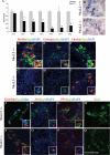
FIGURE 1.
FIGURE 1.
NeuroD1 expression is decreased in the Nkx2.2 null pancreas. Shown is quantitative PCR analysis of NeuroD1 mRNA levels at successive developmental time points during pancreas organogenesis. Nkx2.2−/− levels (black bars) are represented by -fold change in comparison to wild type (A). Statistical significance determined with a Student t test comparing each individual embryonic age group; *, p value<0.05. Error bars represent ±S.E. RNA in situ hybridization comparing NeuroD1 mRNA expression in e15.5 wild-type (B) and Nkx2.2−/− pancreata (C). Immunofluorescence staining of embryonic pancreata in e16.5 Nkx2.2+/−;NeuroD1+/lacZ (D–F and J–M) and Nkx2.2−/−;NeuroD1+/lacZ (G–I and N–Q). NeuroD1 expression is represented by β-galactosidase staining (green, D–Q). Insulin (red, D and G), glucagon (red, E and H; blue, J and N), ghrelin (red, F, I, J, and N), PP (red, L and P), and somatostatin (red, K and O) are shown (confocal images: magnification, 45×; insets: magnification, 90×). β-Galactosidase expression in the Nkx2.2 null tissue was captured with a gain setting 3-fold above control samples to demonstrate expression is maintained, but at decreased levels (compare Q to M).
FIGURE 2.
FIGURE 2.
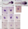
Nkx2.2 regulation of NeuroD1 expression is conserved in zebrafish. In situ hybridization of wild-type 48-hours postfertilization zebrafish embryos for Nkx2.2a (A, B, and B′). In situ hybridization of 48-hours postfertilization zebrafish embryonic pancreas for NeuroD1 (C–E) showing a decrease in NeuroD1 expression in Nkx2.2 splice morphant embryos (E) and either wild-type (C) or control morphant embryos (D).
FIGURE 3.
FIGURE 3.
Nkx2.2 regulates endogenous NeuroD1 and a minimal NeuroD1 promoter. βTC6 cells were transduced with adenovirus containing an siRNA(283) directed against Nkx2.2. Endogenous Nkx2.2 mRNA expression levels (A) and NeuroD1 mRNA expression levels (B) were measured at 48 and 72 h post transduction. Values were normalized to cyclophilin B. Asterisks denote statistically significant decreases between the treated and untreated samples as assessed by Student's t test: *, p < 0.05. Panc1 cells were transfected with either pGL3B vector alone or pGL3B containing the 2.2-kb NeuroD1 promoter and Nkx2.2, the Nkx2.2 binding site mutant (Nkx2.2DBDmut), Ngn3, or Nkx2.2 and Ngn3, in combination (C). Sites are represented by circles, and E-box elements by are represented by rectangles. Values are normalized to Renilla luciferase. Asterisks denote statistically significant changes in luciferase activity upon addition of a regulatory factor as assessed by a student t test: **, p < 0.01; ##, p < 0.01 for Nkx2.2 compared with Nkx2.2DBDmut. Alterations in endogenous NeuroD1 mRNA levels were determined (D). Nkx2.2 protein (G) and mRNA (E) levels were assessed, as were Ngn3 mRNA levels (F). Asterisks denote statistically significant changes in expression levels: *, p < 0.05. Formaldehyde cross-linked e13.5 chromatin was incubated with antibodies specific to Nkx2.2. Immunoprecipitated DNA was analyzed by PCR with primers specific to NeuroD1, insulin, and MafA (H). For controls, PCR was performed with total input DNA (1/10 dilution, Input) and DNA immunoprecipitated with mouse IgG (mIgG). Ratios between ChIP and 10% input were compared between promoter primer sets in the ChIP experiment shown here (I).
FIGURE 4.
FIGURE 4.
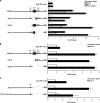
Nkx2.2 activates two separate regions of the NeuroD1 promoter. Panc1 cells were transfected with the full-length 2.2-kb NeuroD1 promoter region or NeuroD1 promoter deletion constructs and Nkx2.2, Ngn3, or Nkx2.2 and Ngn3, in combination. The NeuroD1 promoter constructs are denoted as follows: (A–C) NDfull (2.2 kb (21)), (A) NDΔ1 (deletion between −686 and −240 bp), NDΔ2 (deletion between −2187 and −686 bp), NDΔ3 (deletion between −240 and −113 bp), (B) NDΔ4 (deletion between −2187 and −240 bp), (B, C) NDΔ5 (deletion between −686 and −113 bp), and (C) NDΔ5-Nk1mut (lacking the Nkx2.2 consensus site). Potential NKx2.2 binding sites are represented by circles, and E boxes are represented by rectangles. Statistical analysis was performed by using Student's t tests comparing addition of transcription factor(s) to promoter-alone values. **, p value < 0.01.
FIGURE 5.
FIGURE 5.
Two Nkx2.2-regulated NeuroD1 promoter regions are important for maximal activity in β cells. βTC6 cells were transfected with luciferase promoter constructs NDfull, NDΔ1–5, and two constructs that harbored Site 1 consensus core sequence deletions: NDfull-Nk1mut and NDΔ5-Nk1mut. Potential NKx2.2 binding sites are represented by circles, and E boxes are represented by rectangles. Statistical analysis was performed by using Student's t tests comparing each construct to NDfull activity or between bracketed constructs. *, p value < 0.05.
FIGURE 6.
FIGURE 6.
Nkx2.2 directly binds Site 1 DNA in the distal NeuroD1 promoter. The positive Nkx2.2 binding control reaction with known consensus probe (NkBDctl) was assessed for nuclear extract and in vitro Nkx2.2 binding (A). Nkx2.2 protein binding to consensus site Site 1 (B) was assessed by EMSA analysis performed with in vitro translated Nkx2.2 (lane 3) or DNA binding mutant Nkx2.2 (lane 2). Specificity of any protein-DNA complex formed from βTC6 nuclear extract (lanes 4 and 5) was assayed by incubation with anti-Nkx2.2 antibody (lane 5). The Nkx2.2 antibody incubation resulted in a supershift of the Nkx2.2 containing complex (lane 5). TNT, in vitro translated; NE, nuclear extract.

References

    1. Pictet R., Rutter W. J. (1972) Development of Embryonic Endocrine Pancreas, pp. 26–28, Williams & Wilkins, Washington, DC
    1. Murtaugh L. C. (2007) Development 134, 427–438 - PubMed
    1. Jørgensen M. C., Ahnfelt-Rønne J., Hald J., Madsen O. D., Serup P., Hecksher-Sørensen J. (2007) Endocr. Rev. 28, 685–705 - PubMed
    1. Collombat P., Hecksher-Sørensen J., Serup P., Mansouri A. (2006) Mech. dev. 123, 501–512 - PubMed
    1. Habener J. F., Kemp D. M., Thomas M. K. (2005) Endocrinology 146, 1025–1034 - PubMed

Publication types

MeSH terms