. 2009 Dec;83(23):12101-7.
doi: 10.1128/JVI.01637-09. Epub 2009 Sep 16.

Association of the pr peptides with dengue virus at acidic pH blocks membrane fusion

Affiliations

Association of the pr peptides with dengue virus at acidic pH blocks membrane fusion

I-M Yu et al. J Virol. 2009 Dec.

Abstract

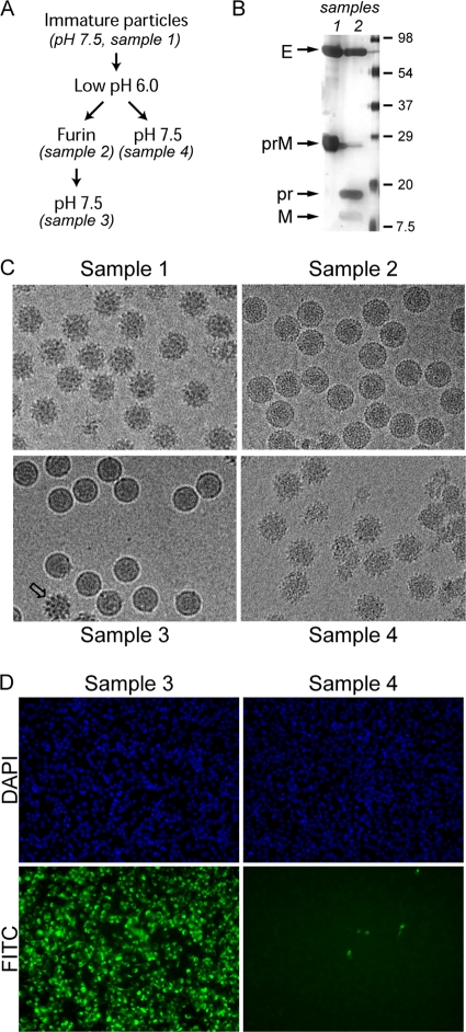
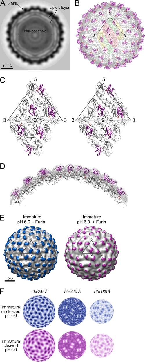
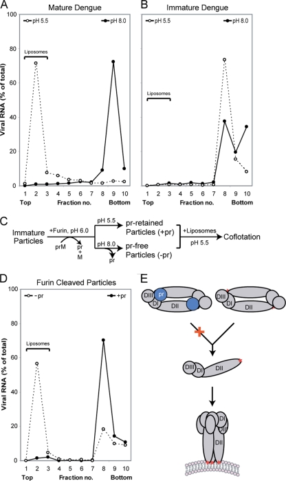
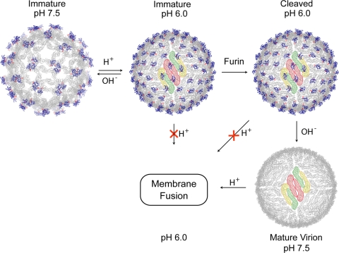
Flavivirus assembles into an inert particle that requires proteolytic activation by furin to enable transmission to other hosts. We previously showed that immature virus undergoes a conformational change at low pH that renders it accessible to furin (I. M. Yu, W. Zhang, H. A. Holdaway, L. Li, V. A. Kostyuchenko, P. R. Chipman, R. J. Kuhn, M. G. Rossmann, and J. Chen, Science 319:1834-1837, 2008). Here we show, using cryoelectron microscopy, that the structure of immature dengue virus at pH 6.0 is essentially the same before and after the cleavage of prM. The structure shows that after cleavage, the proteolytic product pr remains associated with the virion at acidic pH, and that furin cleavage by itself does not induce any major conformational changes. We also show by liposome cofloatation experiments that pr retention prevents membrane insertion, suggesting that pr is present on the virion in the trans-Golgi network to protect the progeny virus from fusion within the host cell.


Figures

FIG. 1.
FIG. 1.
Maturation of dengue immature particles by in vitro furin cleavage. (A) Flow chart of pH and furin treatments of prM-containing particles. (B) Samples 1 and 2 were analyzed by SDS-PAGE silver staining to examine the extent of furin cleavage. The capsid protein was not visible by silver stain, likely due to the absence of cysteine residues (6). (C) EM of the virus particles subjected to pH changes and furin treatment as outlined for panel A. (D) The infectivity of low-pH-treated particles with and without furin cleavage was analyzed by an immunofluorescence assay. Virus-infected BHK cells were probed with a monoclonal anti-E antibody and detected using a fluorescein isothiocyanate (FITC)-conjugated secondary antibody. Nuclei of all cells were counterstained with 4′,6′-diamidino-2-phenylindole (DAPI). The cells were visualized with an epifluorescent microscope. The samples were infected with the same amount of virus particles.
FIG. 2.
FIG. 2.
Structure of furin-cleaved immature virus at pH 6.0. (A) A central cross-section viewed along an icosahedral threefold axis. The darkness of the shading is proportional to the magnitude of the cryoEM electron density. Viral components are labeled. (B) Pseudoatomic structure of the virion upon fitting the crystal structures of prM-E into the cryoEM map. The E and pr proteins are shown in gray and magenta, respectively. The fusion loop is colored in red. An icosahedral asymmetry unit is outlined in a triangle. The E dimer on the icosahedral twofold axis is shaded pink, whereas the two monomers of the general-position dimer are shaded yellow and green. (C) Stereo diagram showing the interactions of pr (magenta) with E (gray). (D) A cross-section through the cryoEM density showing the overall quality of the fitting. The EM density is shown in gray, and protein molecules are colored as described for panel C. (E) Structure comparison of the immature virus with and without furin cleavage. The difference density calculated by subtracting the density of the mature virion is shaded in color (blue, before furin cleavage; magenta, after furin cleavage). An icosahedral asymmetric unit is outlined in a black triangle. (F) Comparison of the radial density sections of the immature particles with and without furin treatment. Maps at radii corresponding to the outer shell (r1), inner shell (r2), and membrane region (r3), respectively, are shown.
FIG. 3.
FIG. 3.
Association of pr inhibits membrane insertion. Virus particles were mixed with liposomes at pH 5.5 and analyzed by sedimentation in sucrose gradients. Fractions from the gradients were analyzed by qRT-PCR to determine the amount of viral RNA. Liposomes float to the top of the gradient, whereas virus particles sediment to the bottom. (A) Mature dengue virus harvested from infected cells. (B) Immature dengue particles. (C) Flow chart of in vitro furin treatment for samples used in panel D. (D) Furin-treated particles with (−pr) and without (+pr) neutralization. (E) Conformational changes of the glycoproteins necessary to promote membrane fusion. E proteins are shown in gray, and the three domains are labeled (DI, DII, and DIII). The fusion loops are indicated by a red star. The presence of pr (blue) likely prevents E dimer dissociation at low pH.
FIG. 4.
FIG. 4.
Conformational metamorphosis of dengue viruses. Structures of the virion at different stages of the life cycle are shown. The E and pr proteins are shown in gray and blue, respectively, and the fusion loops are in red. An E protein raft, consisting of three parallel dimmers, is shaded in color.

References

    1. Allison, S. L., J. Schalich, K. Stiasny, C. W. Mandl, C. Kunz, and F. X. Heinz. 1995. Oligomeric rearrangement of tick-borne encephalitis virus envelope proteins induced by an acidic pH. J. Virol. 69:695-700. - PMC - PubMed
    1. Baker, T. S., and R. H. Cheng. 1996. A model-based approach for determining orientations of biological macromolecules imaged by cryoelectron microscopy. J. Struct. Biol. 116:120-130. - PubMed
    1. Baker, T. S., N. H. Olson, and S. D. Fuller. 1999. Adding the third dimension to virus life cycles: three-dimensional reconstruction of icosahedral viruses from cryo-electron micrographs. Microbiol. Mol. Biol. Rev. 63:862-922. - PMC - PubMed
    1. Bressanelli, S., K. Stiasny, S. L. Allison, E. A. Stura, S. Duquerroy, J. Lescar, F. X. Heinz, and F. A. Rey. 2004. Structure of a flavivirus envelope glycoprotein in its low-pH-induced membrane fusion conformation. EMBO J. 23:728-738. - PMC - PubMed
    1. Chen, J., K. H. Lee, D. A. Steinhauer, D. J. Stevens, J. J. Skehel, and D. C. Wiley. 1998. Structure of the hemagglutinin precursor cleavage site, a determinant of influenza pathogenicity and the origin of the labile conformation. Cell 95:409-417. - PubMed

Publication types