. 2009 Dec;83(23):12399-406.
doi: 10.1128/JVI.00888-09. Epub 2009 Sep 16.

Control of herpes simplex virus replication is mediated through an interferon regulatory factor 3-dependent pathway

Affiliations

Control of herpes simplex virus replication is mediated through an interferon regulatory factor 3-dependent pathway

Vineet D Menachery et al. J Virol. 2009 Dec.

Abstract

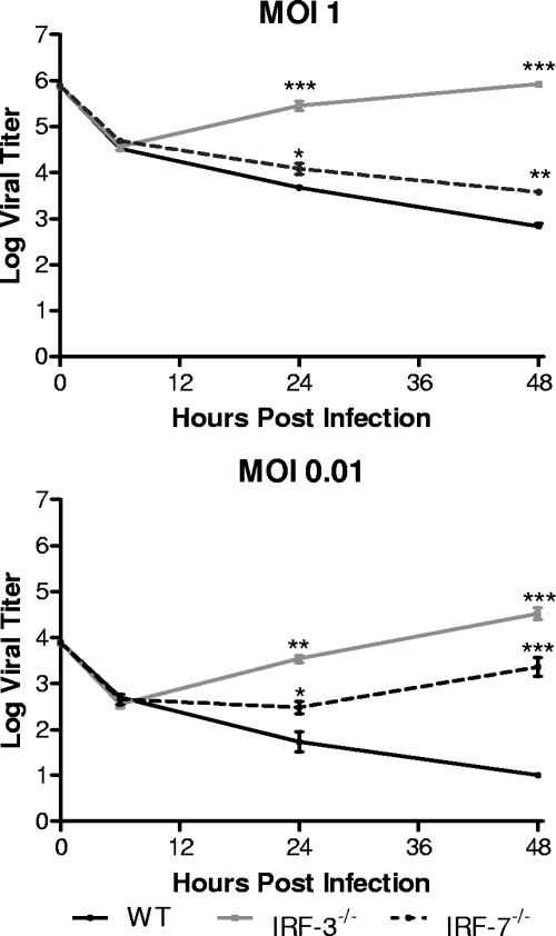
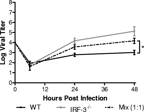
The type I interferon (IFN) cascade is critical in controlling viral replication and pathogenesis. Recognition pathways triggered by viral infection rapidly induce the type I IFN cascade, often in an IFN regulatory factor 3 (IRF-3)-dependent fashion. This dependence predicts that loss of IRF-3 would render early recognition pathways inoperative and thereby impact virus replication, but this has not been observed previously with herpes simplex virus type 1 (HSV-1) in vitro. In this study, HSV-1-infected IRF-3(-/-) bone marrow-derived dendritic cells (BMDCs) and macrophages supported increased HSV replication compared to control cells. In addition, IRF-3-deficient BMDCs exhibited delayed type I IFN synthesis compared to control cells. However, while IFN pretreatment of IRF-3(-/-) BMDCs resulted in reduced virus titers, a far greater reduction was seen after IFN treatment of wild-type cells. This suggests that even in the presence of exogenously supplied IFN, IRF-3(-/-) BMDCs are inherently defective in the control of HSV-1 replication. Together, these results demonstrate a critical role for IRF-3-mediated pathways in controlling HSV-1 replication in cells of the murine immune system.


Figures

FIG. 1.
FIG. 1.
In vitro replication in bone marrow derived dendritic cells. Primary BMDCs were infected with WT HSV-1 at an MOI of 1 or 0.01. At indicated times postinfection, cells and supernatants were harvested and virus titers assayed on Vero cells. The results shown are the mean titers of three independent experiments. *, P < 0.05; **, P < 0.01.
FIG. 2.
FIG. 2.
In vitro replication in bone marrow derived macrophages. Primary BMM were infected with WT HSV-1 at an MOI of 1 or 0.01. At the indicated times postinfection, cells and supernatants were harvested, and virus titers were assayed on Vero cells. The results shown are mean titers of four independent experiments. *, P < 0.05; **, P < 0.01; ***, P < 0.001.
FIG. 3.
FIG. 3.
In vitro replication in BMDCs lacking IFN signaling. Primary BMDCs derived from WT and IFN-α/β/γ receptor-deficient (AG129) or STAT1-deficient mice were infected with WT HSV-1 at an MOI of 1 or 0.01. At indicated times postinfection, cells and supernatants were harvested, and virus titers were assayed on Vero cells. The results shown are the mean titers of three independent experiments. *, P < 0.05; **, P < 0.01; ***, P < 0.001.
FIG. 4.
FIG. 4.
IFN-β secretion by infected BMDCs. Primary BMDCs were infected with WT HSV-1 at an MOI of 5. At indicated times postinfection, cells and supernatants were harvested. Cells were removed by low-speed centrifugation, and supernatants were assayed for IFN-β by ELISA. The results in the upper panel are mean totals from three independent experiments. Cells and supernatants were also assayed for virus titers at 6, 12, and 24 h postinfection, and virus titers were assayed on Vero cells. The results in the lower panel shown are the mean titers of three independent experiments. *, P < 0.05.
FIG. 5.
FIG. 5.
IFNαR blockade in BMDCs. Primary BMDCs were infected with WT HSV-1 at an MOI of 0.01. After infection, BMDCs were plated in media containing 5 μg of IFNαR blocking antibody (MAR1-5A3)/ml for the duration of the experiment. At the indicated times postinfection, cells and supernatants were harvested, and virus titers were assayed on Vero cells. The results shown are the mean titers of three independent experiments. *, P <, 0.05; **, P < 0.01.
FIG. 6.
FIG. 6.
In vitro replication after mixing BMDC populations. Primary WT and IRF-3−/− BMDCs were mixed at a 1:1 ratio and infected at an MOI of 0.01. At the indicated times postinfection, cells and supernatants were harvested, and virus titers were assayed on Vero cells. The results shown are the mean titers of three independent experiments. *, P < 0.05.
FIG. 7.
FIG. 7.
In vitro replication after IFN-β pretreatment of BMDCs. Primary WT and IRF-3−/− BMDCs were pretreated with 100 U of mouse IFN-β/ml for 16 h. Cells were then infected with WT HSV-1 at an MOI of 0.01. At the indicated times postinfection, cells and supernatants were harvested, and virus titers were assayed on Vero cells. The results shown are the mean titers of three independent experiments. *, P < 0.05; **, P < 0.01.
FIG. 8.
FIG. 8.
Model for continued defect in IRF-3-deficient BMDCs. Postattachment, HSV-1 infection is recognized through an unknown sensor mechanism that leads to activation of IRF-3. The early recognition pathway mediates the production of type I IFN- and IRF-3-dependent ISGs, leading to the control of HSV-1 replication via the type I IFN cascade. However, pretreatment with IFN does not restore HSV-1 replication in IRF-3−/− BMDCs to WT levels. The continued defect is potentially due to three, nonexclusive mechanisms outlined in white squares: defective IFN amplification, defective antiviral trigger signaling, and IRF-3-dependent gene synergy with the antiviral response. One or more of these mechanisms leads to continued defect in the control of HSV-1 replication in IRF-3−/− BMDCs compared to WT BMDCs after IFN treatment.

References

    1. Andersen, J., S. VanScoy, T. F. Cheng, D. Gomez, and N. C. Reich. 2008. IRF-3-dependent and augmented target genes during viral infection. Genes Immun. 9:168-175. - PubMed
    1. Bauer, D., S. Mrzyk, N. van Rooijen, K. P. Steuhl, and A. Heiligenhaus. 2000. Macrophage-depletion influences the course of murine HSV-1 keratitis. Curr. Eye Res. 20:45-53. - PubMed
    1. Bowie, A. G., and L. Unterholzner. 2008. Viral evasion and subversion of pattern-recognition receptor signalling. Nat. Rev. Immunol. 8:911-922. - PMC - PubMed
    1. Cella, M., D. Jarrossay, F. Facchetti, O. Alebardi, H. Nakajima, A. Lanzavecchia, and M. Colonna. 1999. Plasmacytoid monocytes migrate to inflamed lymph nodes and produce large amounts of type I interferon. Nat. Med. 5:919-923. - PubMed
    1. Cheng, H., T. M. Tumpey, H. F. Staats, N. van Rooijen, J. E. Oakes, and R. N. Lausch. 2000. Role of macrophages in restricting herpes simplex virus type 1 growth after ocular infection. Investig. Ophthalmol. Vis. Sci. 41:1402-1409. - PubMed

Publication types

Substances

LinkOut - more resources