. 2009 Dec;83(23):12473-82.
doi: 10.1128/JVI.01138-09. Epub 2009 Sep 16.

Identification and characterization of broadly neutralizing human monoclonal antibodies directed against the E2 envelope glycoprotein of hepatitis C virus

Affiliations

Identification and characterization of broadly neutralizing human monoclonal antibodies directed against the E2 envelope glycoprotein of hepatitis C virus

Teresa J Broering et al. J Virol. 2009 Dec.

Abstract

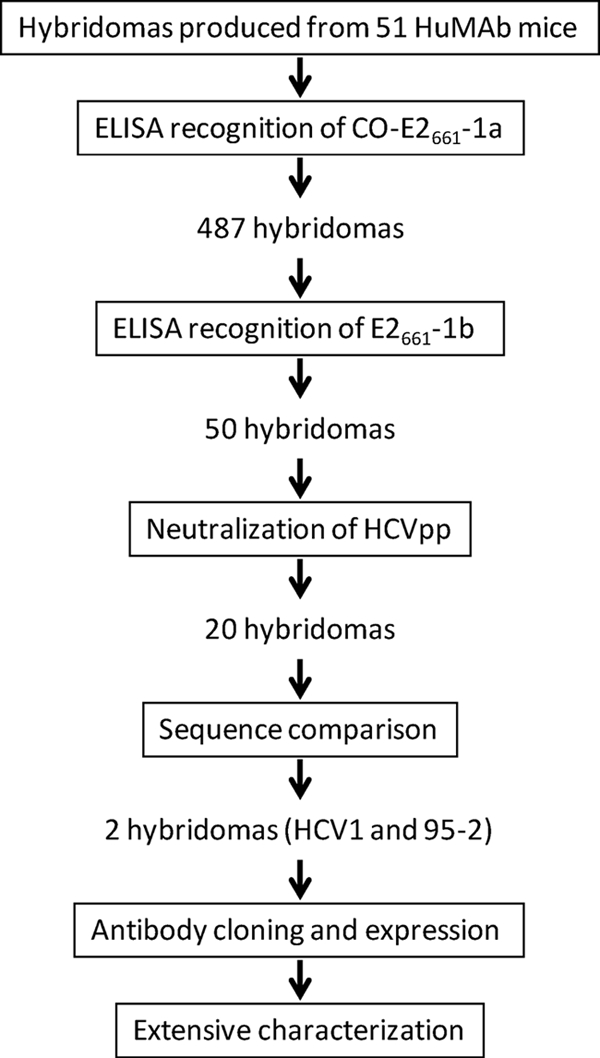
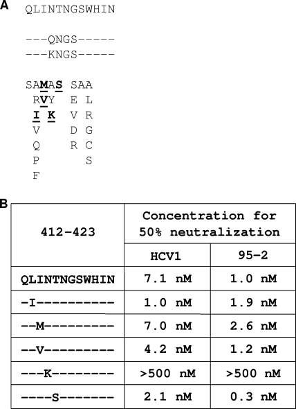
Nearly all livers transplanted into hepatitis C virus (HCV)-positive patients become infected with HCV, and 10 to 25% of reinfected livers develop cirrhosis within 5 years. Neutralizing monoclonal antibody could be an effective therapy for the prevention of infection in a transplant setting. To pursue this treatment modality, we developed human monoclonal antibodies (HuMAbs) directed against the HCV E2 envelope glycoprotein and assessed the capacity of these HuMAbs to neutralize a broad panel of HCV genotypes. HuMAb antibodies were generated by immunizing transgenic mice containing human antibody genes (HuMAb mice; Medarex Inc.) with soluble E2 envelope glycoprotein derived from a genotype 1a virus (H77). Two HuMAbs, HCV1 and 95-2, were selected for further study based on initial cross-reactivity with soluble E2 glycoproteins derived from genotypes 1a and 1b, as well as neutralization of lentivirus pseudotyped with HCV 1a and 1b envelope glycoproteins. Additionally, HuMAbs HCV1 and 95-2 potently neutralized pseudoviruses from all genotypes tested (1a, 1b, 2b, 3a, and 4a). Epitope mapping with mammalian and bacterially expressed proteins, as well as synthetic peptides, revealed that HuMAbs HCV1 and 95-2 recognize a highly conserved linear epitope spanning amino acids 412 to 423 of the E2 glycoprotein. The capacity to recognize and neutralize a broad range of genotypes, the highly conserved E2 epitope, and the fully human nature of the antibodies make HuMAbs HCV1 and 95-2 excellent candidates for treatment of HCV-positive individuals undergoing liver transplantation.


Figures

FIG. 1.
FIG. 1.
Schematic representation of rapid screening method devised to select lead-candidate hybridomas. The large panel of CO-E2661-1a-reactive hybridomas were screened for reactivity with E2661-1b as well as for neutralization of genotype 1a HCVpp. Cross-reactive, neutralizing antibodies produced by the hybridomas were sequenced and grouped according to both amino acid identity and similarity in VH gene usage. HCV1 and 95-2 were selected as lead candidates, cloned into expression vectors, and transfected into CHO cells for the production of recombinant antibody. Antibodies were purified and subsequently characterized more extensively.
FIG. 2.
FIG. 2.
CDRs of broadly cross-reactive lead-candidate HuMAbs. CDR1, CDR2, and CDR3 of the four unique sequences of cross-reactive anti-HCV HuMAbs are shown, with alignment scores denoted below the amino acid entries. 95-14, 95-38, and 95-2 were isolated from mouse 97895, and HCV1 was isolated from mouse 85083.
FIG. 3.
FIG. 3.
HuMAbs HCV1 and 95-2 interact with HCV E2 glycoproteins derived from genotypes 1a and 1b. HuMAbs HCV1 (A) and 95-2 (B) and mouse His6-directed antibody (C) were tested for the capacity to bind to soluble E2 glycoproteins derived from genotypes 1a and 1b in ELISA. ELISA plates were coated with 2 μg/ml of CO-E2661-1a (triangles) or E2661-1b (squares), and a dilution series of each antibody was introduced to the assay, as well as the mouse MAb directed against the His6 epitope tag present in each E2 protein. Bound antibody was detected employing goat anti-human secondary antibody (HuMAbs) or goat anti-mouse secondary antibody (mouse anti-His6) conjugated to AP, followed by development with PNPP substrate, and the absorbance at 405 nm for each dilution was plotted.
FIG. 4.
FIG. 4.
HCV1 and 95-2 neutralize HCVpp representing a diverse group of HCV genotypes. Fivefold dilutions of HuMAbs HCV1 (squares) and 95-2 (diamonds) and an irrelevant HuMAb (negative control; triangles) were incubated for 1 h at room temperature with lentiviral pseudovirus with an engineered luciferase gene. The pseudovirus (HCVpp) was generated with E1/E2 glycoproteins from the following genotypes: H77 1a (A), GR5 1a (B), 1b (C), 2b (D), 3a (E), and 4a (F). The HCVpp-antibody mixture was added to Hep3B cells, followed by incubation at 37°C for 72 h. Infection was assessed with Brightglo luciferase reagent, followed by analysis using a Victor3 multilabel reader. Light output, expressed as counts per second (CPS), was plotted.
FIG. 5.
FIG. 5.
HuMAbs HCV1 and 95-2 recognize amino acids 412 to 423 of the E2 envelope glycoprotein. The top graphic depicts the amino-terminal 80 amino acids of the E2 envelope glycoprotein, with the amino acid numbers indicated above the schematic. Hypervariable region 1 (HVR1) is denoted with hashing. Various bacterial fusion proteins representing different truncation protein products of the E2 glycoprotein were generated. The corresponding amino acids for each truncated E2 product are indicated. The various truncations were assayed in ELISA for recognition by HuMAbs HCV1 and 95-2. Bound antibodies were detected with goat anti-human antibody conjugated to AP, followed by PNPP substrate addition. ELISA results are listed to the right of the schematic; positive recognition is indicated by a plus sign, while signals equivalent to background are indicated by a minus sign.
FIG. 6.
FIG. 6.
Determination of critical residues required for HCV1 and 95-2 binding. (A) The sequence of the 412-423 region of E2 is listed in one-letter amino acid code for the genotype 1a H77 strain that was used for HCVpp generation. Listed below that sequence are the sequences for all strains of HCVpp that were tested in Fig. 4. Positions identical to the position in H77 are indicated by dashes, while differences are indicated in one-letter amino acid code. (B) Bacterially expressed fusion proteins containing E2 amino acids 412 to 423 with the indicated amino acid mutated to alanine were assayed in ELISA with 0.052 nM HuMAbs HCV1 and 95-2. Bound antibodies were detected with goat anti-human antibody conjugated to AP. The data were plotted as percentages of the absorbance obtained with the wild-type Trx-E2412-423-His protein derived from native H77.
FIG. 7.
FIG. 7.
HCV1 and 95-2 bind E2 amino acids 412 to 423 isolated from viruses of infected patients, representing a diverse group of genotypes. (A) Bacterially expressed fusion proteins containing E2 amino acids 412 to 423 of isolates identified from the NIH HCV database (y axis) were used to coat ELISA plates and probed with 0.026 nM HuMAbs HCV1 (black) and 95-2 (gray). The top-most sequence represents the H77 genotype 1a 412-423 sequence. In subsequent listings, positions identical to the position in H77 are indicated by dashes, and differences are listed in one-letter amino acid code. Absorbance was plotted on the x axis. Various dilutions of HuMAbs HCV1 (B) and 95-2 (C) were measured in ELISA against the E2 412-423 fusion proteins that gave suboptimal absorbance in panel A. Each assayed protein, with the corresponding amino acid sequence and symbol, is listed in the legend. As a control for maximal antibody binding kinetics, the fusion protein containing amino acids 412 to 423 of the H77 1a sequence (squares) was included. Absorbance was plotted as a function of antibody concentration.
FIG. 8.
FIG. 8.
Generation and neutralization of HCVpp containing E2 envelope glycoproteins with mutations in amino acids 412 to 423. (A) A total of 28 mutations were introduced into amino acids 412 to 423 of the genotype 1a (H77) full-length E2 envelope glycoprotein (listed at the top), using site-directed mutagenesis. Directly below are listed two mutants that had four amino acid changes each from the H77 sequence. The other 26 mutants had single amino acid changes and are listed in the lower part of the panel. The five bold, underlined amino acid changes represent mutations that yielded infectious HCVpp. Unchanged amino acids are indicated as dashes, and the altered positions are indicated with the one-letter amino acid code. (B) Fivefold dilutions of HuMAbs HCV1 and 95-2 were incubated for 1 h at room temperature with HCVpp generated with H77 1a E1/E2 glycoproteins or H77 1a E1/E2 glycoproteins containing the desired changes in the E2 protein, indicated to the left. The neutralization assays were performed and developed as described in the legend to Fig. 4. The concentration of antibody required to reduce the light output to 50% of that of the negative control antibody is indicated for both HCV1 and 95-2.

References

    1. Allander, T., K. Drakenberg, A. Beyene, D. Rosa, S. Abrignani, M. Houghton, A. Widell, L. Grillner, and M. A. Persson. 2000. Recombinant human monoclonal antibodies against different conformational epitopes of the E2 envelope glycoprotein of hepatitis C virus that inhibit its interaction with CD81. J. Gen. Virol. 81:2451-2459. - PubMed
    1. Babcock, G. J., T. J. Broering, H. J. Hernandez, R. B. Mandell, K. Donahue, N. Boatright, A. M. Stack, I. Lowy, R. Graziano, D. Molrine, D. M. Ambrosino, and W. D. Thomas, Jr. 2006. Human monoclonal antibodies directed against toxins A and B prevent Clostridium difficile-induced mortality in hamsters. Infect. Immun. 74:6339-6347. - PMC - PubMed
    1. Babcock, G. J., D. J. Esshaki, W. D. Thomas, Jr., and D. M. Ambrosino. 2004. Amino acids 270 to 510 of the severe acute respiratory syndrome coronavirus spike protein are required for interaction with receptor. J. Virol. 78:4552-4560. - PMC - PubMed
    1. Bartosch, B., J. Dubuisson, and F. L. Cosset. 2003. Infectious hepatitis C virus pseudo-particles containing functional E1-E2 envelope protein complexes. J. Exp. Med. 197:633-642. - PMC - PubMed
    1. Bugli, F., N. Mancini, C. Y. Kang, C. Di Campli, A. Grieco, A. Manzin, A. Gabrielli, A. Gasbarrini, G. Fadda, P. E. Varaldo, M. Clementi, and R. Burioni. 2001. Mapping B-cell epitopes of hepatitis C virus E2 glycoprotein using human monoclonal antibodies from phage display libraries. J. Virol. 75:9986-9990. - PMC - PubMed

Substances