. 2009 Nov;83(22):11655-64.
doi: 10.1128/JVI.01040-09. Epub 2009 Sep 16.

Adeno-associated virus site-specific integration is mediated by proteins of the nonhomologous end-joining pathway

Affiliations

Adeno-associated virus site-specific integration is mediated by proteins of the nonhomologous end-joining pathway

Shyam Daya et al. J Virol. 2009 Nov.

Abstract

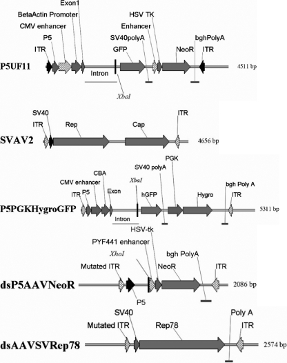
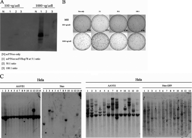
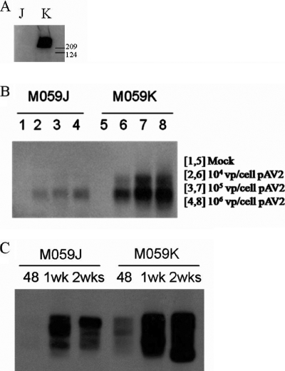
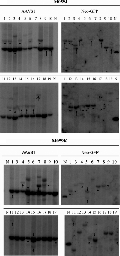
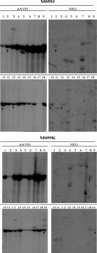
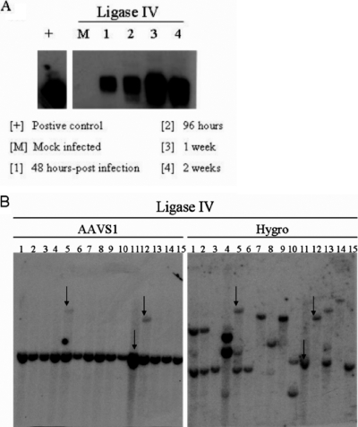
Adeno-associated virus type 2 (AAV 2) is the only eukaryotic virus capable of site-specific integration; the target site is at chromosome 19q13.4, a site termed AAVS1. The biology of AAV latency has been extensively studied in cell culture, yet the precise mechanism and the required cellular factors are not known. In this study, we assessed the relative frequencies of stable site-specific integration by characterization of cell clones containing integrated AAV vectors. By this assay, two proteins involved in nonhomologous end joining (NHEJ), DNAPKcs and ligase IV, exhibit differential effects on AAV site-specific integration. DNAPKcs is not required; its presence increases the frequency of junction formation indicative of site-specific integration, but seems to reduce the ratio of site-specific integration to random integration (i.e., the latter is even more enhanced). In contrast, site-specific integration is significantly reduced relative to random integration in cells deficient in ligase IV expression. Furthermore, we show that single-stranded AAV vectors are better substrates for site-specific integration than are self-complementary AAV vectors; the absence of DNAPKcs did not affect the targeted integration of these double-stranded AAV vectors. Together, these data suggest that NHEJ proteins participate in site-specific integration, and indicate a role for the single-stranded form of AAV DNA in targeted integration.


Figures

FIG. 1.
FIG. 1.
Schematic representation of vector constructs. The single-stranded AAV vectors include P5UF11, SVAV2, and P5PGKHygroGFP. The self-complementary AAV vectors include dsP5AAVNeoR and dsAAVSVRep78. The important restriction sites (XbaI, XhoI) used for Southern analysis are marked. There are no EcoRV restriction sites in these vectors.
FIG. 2.
FIG. 2.
scAAV vectors integrate site-specifically in HeLa cells. (A) Junction PCR assay of HeLa cells infected with dsP5AAVNeoR only or with dsAAV-SVRep78 at different ratios and MOIs (viral genomes per cell [vg/cell]) analyzed 1 week postinfection. (B) Colony-forming assay. Ten thousand infected HeLa cells were seeded into 100 mM dishes and selected with G418 for 2 weeks. Postselection, the resistant colonies were stained with crystal violet and counted. The counts are shown only for the lower dose (100 vg/cell). Visually, more colonies are seen at the 5:1 ratio at both doses. (C) Comparison of ssAAV and scAAV specific integration. Left panel, HeLa scAAV clones (50:1; dsP5AAVNeoR:dsAAVSVRep78; MOI, 105). Right panel, HeLa ssAAV clones (50:1; P5UF11:SVAV2; MOI, 105). The arrows indicate the clones that have both AAVS1 and AAV signals colinked.
FIG. 3.
FIG. 3.
AAV-AAVS1 junction product formation in M059J and M059K cells. (A) Western blot for DNAPKcs from whole-cell extracts from M059J (J) and M059K (K) cells. Sizes in kilodaltons are marked on the side. DNAPKcs is approximately 460 kDa in size. (B) Junction formation at different doses of wild-type AAV 2 infection. The cell lines were infected with different doses (number of viral particles [vp] per cell) of wild-type AAV 2. Genomic DNA from infected cells was isolated 48 h postinfection for junction assay. (C) Time course of junction formation. Both cells were infected with 106 viral genomes per cell (50:1 ratio of P5UF11 to SVAV2). Genomic DNA was isolated at 48 h, 1 week, or 2 weeks postinfection for junction assay.
FIG. 4.
FIG. 4.
Southern hybridization of M059J and M059K clones infected with single-stranded AAV vectors P5UF11 and SVAV2 (50:1 ratio, 106 viral particles [vp]/cell). Representative blots are shown. The left blots show signals for AAVS1 sequences, and the right blots show signals for neomycin. The arrows indicate the clones that had both AAVS1 and neomycin signals colinked. The endogenous AAVS1 band is seen in all clones. The presence of additional AAVS1 bands is indicative of rearrangements of the site, which occurs when it is targeted. In some cases, the endogenous AAVS1 is colinked with AAV, indicative of deletions occurring before integration. If integration occurs with minimal deletions, a higher AAVS1 band colinks with AAV. The samples are numbered. N represents a positive control plasmid which was used to determine specificity of the neomycin probe.
FIG. 5.
FIG. 5.
Southern hybridization of M059J and M059K clones infected with self-complementary vectors dsP5AAVNeoR and dsAAVSVRep78 (5:1 ratio, 104 viral genomes/cell). Representative blots are shown. The left blots show signals for AAVS1 sequences, and the right blots show signals for neomycin. The arrows indicate the clones that had both AAVS1 and neomycin signals colinked.
FIG. 6.
FIG. 6.
(A) Time course of junction formation in ligase IV hypomorphic cell lines. Cells were infected with wild-type AAV 2 at 105 viral particles/cell. Genomic DNA was isolated at the indicated time points for junction assay. (B) Southern hybridization of ligase IV clones infected with single-stranded AAV vectors P5PGKHygroGFP and SVAV2 (50:1 ratio, 105 vg/cell). Representative blots are shown. The left blots show signals for AAVS1 sequences, and the right blots show signals for hygromycin. The arrows indicate the clones that have both AAVS1 and hygromycin signals colinked.

References

    1. Atchison, R. W., B. C. Casto, and W. M. Hammon. 1965. Adenovirus-associated defective virus particles. Science 149:754-756. - PubMed
    1. Auricchio, A., M. Hildinger, E. O'Connor, G. P. Gao, and J. M. Wilson. 2001. Isolation of highly infectious and pure adeno-associated virus type 2 vectors with a single-step gravity-flow column. Hum. Gene Ther. 12:71-76. - PubMed
    1. Berns, K. I., T. C. Pinkerton, G. F. Thomas, and M. D. Hoggan. 1975. Detection of adeno-associated virus (AAV)-specific nucleotide sequences in DNA isolated from latently infected Detroit 6 cells. Virology 68:556-560. - PubMed
    1. Blacklow, N. R., M. D. Hoggan, and W. P. Rowe. 1968. Serologic evidence for human infection with adenovirus-associated viruses. J. Natl. Cancer Inst. 40:319-327. - PubMed
    1. Carter, B. J. 2005. Adeno-associated virus vectors in clinical trials. Hum. Gene Ther. 16:541-550. - PubMed

Publication types

LinkOut - more resources