. 2009 Sep 15;23(18):2152-65.
doi: 10.1101/gad.1820109.

MicroRNA dynamics in the stages of tumorigenesis correlate with hallmark capabilities of cancer

Affiliations

MicroRNA dynamics in the stages of tumorigenesis correlate with hallmark capabilities of cancer

Peter Olson et al. Genes Dev. .

Abstract

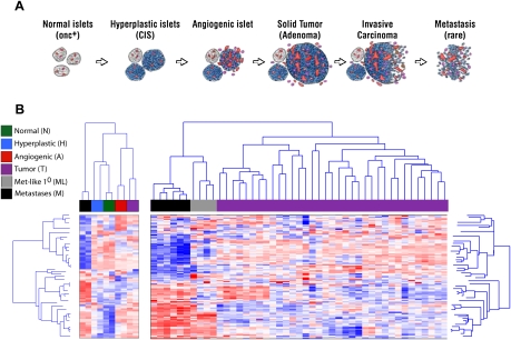
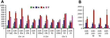
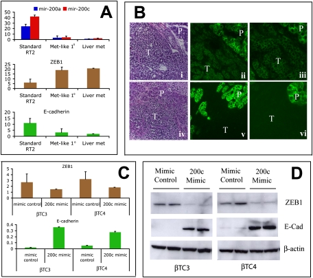
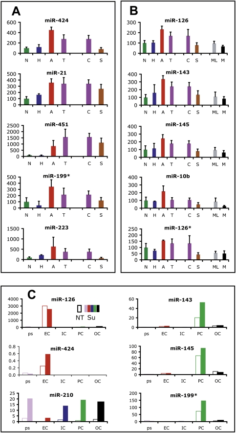
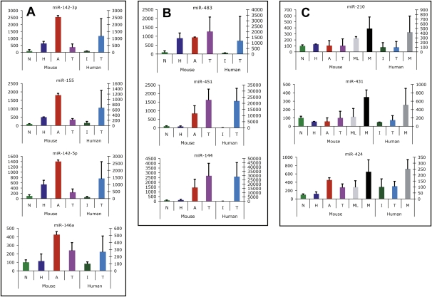
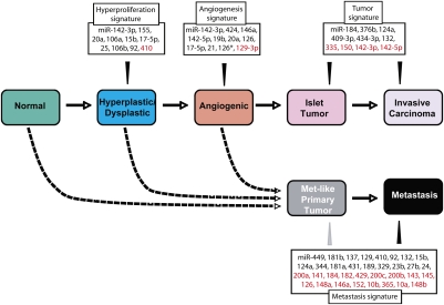
While altered expression of microRNAs (miRs) in tumors has been well documented, it remains unclear how the miR transcriptome intersects neoplastic progression. By profiling the miR transcriptome we identified miR expression signatures associated with steps in tumorigenesis and the acquisition of hallmark capabilities in a prototypical mouse model of cancer. Metastases and a rare subset of primary tumors shared a distinct miR signature, implicating a discrete lineage for metastatic tumors. The miR-200 family is strongly down-regulated in metastases and met-like primary tumors, thereby relieving repression of the mesenchymal transcription factor Zeb1, which in turn suppresses E-cadherin. Treatment with a clinically approved angiogenesis inhibitor normalized angiogenic signature miRs in primary tumors, while altering expression of metastatic signature miRs similarly to liver metastases, suggesting their involvement in adaptive resistance to anti-angiogenic therapy via enhanced metastasis. Many of the miR changes associated with specific stages and hallmark capabilities in the mouse model are similarly altered in human tumors, including cognate pancreatic neuroendocrine tumors, implying a generality.


Figures

Figure 1.
Figure 1.
Differential expression of the miR transcriptome in the distinctive stages of multistep tumorigenesis. (A) Schematic representation of the separable stages of multistep tumorigenesis in the RT2 transgenic mouse model of PNETs, which afforded the experimental approach to miR transcriptome profiling of discrete stages of carcinogenesis. (B) Clustering analysis of normal, hyperplastic, and angiogenic islet pools, along with pools of primary tumors and of metastases, are shown in the left panel. In the right panel, clustering analysis of 39 individual tumors and six liver metastases reveals that a subset of primary tumors is remarkably similar to metastases in its miR expression profile. Another subset of tumors up-regulates miRs in the Dlk1-Gtl2 imprinted cluster (miRs in red two-thirds of the way down in the left-most primary [purple] tumors as well as in metastases; see also Supplemental Fig. 2).
Figure 2.
Figure 2.
miR profiling reveals dynamic regulation and illuminates immune cell infiltration in pretumor stages in RT2 carcinogenesis. (A) Fold changes of the three miR-17-92 clusters by chromosomal location in normal (N), hyperplastic (H), and angiogenic (A) islets, and tumors (T). Average plus standard deviation are shown with expression in normal islets normalized to 100. (B) Expression of miR-150, miR-142-3p, miR-142-5p, and miR-155 in the neoplastic stages. Average plus standard deviation are shown with expression in normal islets normalized to 100%. Supplemental Figure 2 documents the immune cell specificity of these miRs in this pathway.
Figure 3.
Figure 3.
The miR-200–Zeb1–E-cadherin axis is deregulated in metastases and a subset of primary tumors. (A) Expression levels for miR-200a and miR-200c by Q-PCR normalized to an unaffected miR, miR-16. The miR-200 (also known as “miR-8”) family is organized in two clusters in the human and mouse genome. miR-200a and miR-200c are located in separate clusters, and their expression is representative of all miRs from the two clusters. ZEB1 mRNA exhibits a reciprocal expression pattern compared with miR-200 in that it is low in standard RIP-Tag tumors but up-regulated in met-like primary tumors and liver metastases. E-cadherin expression mirrors miR-200 expression and is mutually exclusive with ZEB1. (B) H&E (panels i,iv) and E-cadherin immunofluorescence at 10× (panels ii,v) and 20× (panels iii,vi) magnification for a noninvasive RT2 tumor (panels i–iii) or a highly invasive IC2 tumor (panels iv–vi). (T) Tumor; (P) normal exocrine pancreas. Noninvasive RT2 tumors express E-cadherin, whereas expression is not detected in IC2s. Note the even higher levels of E-cadherin in normal exocrine pancreas as compared with noninvasive tumors, which are nevertheless positive. (C,D) RNA (C) and protein (D) levels of ZEB1 and E-cadherin following electroporation of a miR-200c mimic or mimic control oligos into βTC3 or βTC4 cells demonstrate their regulatory interconnection. Bar graphs show average plus standard deviation.
Figure 4.
Figure 4.
Anti-angiogenic therapy quasinormalizes the angiogenic miR signature, and evokes elements of the metastatic miR signature. (A) miRs up-regulated in angiogenic islets and down-regulated by sunitinib treatment. (B) miRs up-regulated in angiogenic islets, down-regulated by sunitinib treatment, and similarly down-regulated in metastatic lesions. (N) Normal; (H) hyperplastic; (A) angiogenic; (T) tumor; (C) control-treated tumors; (S) sunitinib-treated tumors; (ML) met-like primary tumors; (M) metastases. (C) FACS of the constituent cell types of tumors followed by Q-PCR analysis reveals that affected miRs reflect either the differential abundance of the tumor vasculature and its component endothelial cells and pericytes, or altered regulation of miRs in the predominant population of cancer cells. (Open bars) (NT) Not treated. (Colored bar) (Su) Sunitinib treated; (ps) presort; (EC) endothelial cells; (IC) immune cells; (PC) pericytes; (OC) other cells.
Figure 5.
Figure 5.
miRs similarly regulated in mouse and human PNETs and metastases. (A) miRs differentially expressed between human PNETs and normal human islets, and identified as components of the hyperplastic or angiogenic miR signature in mice. (B) miRs differentially expressed between PNETs and normal human islets and similarly expressed differentially between normal islets and the panel of tumors in mice. (C) miRs differentially regulated between metastases and tumors in both mice and humans. (N) Normal islets; (H) hyperplastic islets; (A) angiogenic islets; (T) tumor; (I) human islets; (ML) met-like primary tumors; (M) liver metastases. Average plus standard deviation are shown, with expression in normal islets normalized to 100%. For C, P-value, average, and standard deviation for tumors, met-like primary tumors, and metastases were determined using values from individual tumors from each group.
Figure 6.
Figure 6.
miR signatures of the stages in multistep tumorigenesis, ascribed to hallmark capabilities. The orchestrated stepwise progression from normality to metastasis during tumorigenesis of the pancreatic islets is marked by distinctive miR signatures of the temporally and histologically separable neoplastic stages, correlating with the acquisition of hallmark capabilities of cancer. In addition, miR profiling uncovered strong correlative evidence that a distinct class of primary tumors spawns the metastases, in contradistinction to expectations of the alternative hypothesis that enabling mutations occur in a few cells of primary tumors, endowing those cells with a metastatic capability. Gray arrowhead from Metastasis signature to Met-like tumor indicates the majority of the metastasis signature is present in met-like primary tumors.

References

    1. Ambros V. The functions of animal microRNAs. Nature. 2004;431:350–355. - PubMed
    1. Bartel DP. MicroRNAs: Genomics, biogenesis, mechanism, and function. Cell. 2004;116:281–297. - PubMed
    1. Bergers G, Hanahan D. Modes of resistance to anti-angiogenic therapy. Natl Rev. 2008;8:592–603. - PMC - PubMed
    1. Bernards R, Weinberg RA. A progression puzzle. Nature. 2002;418:823. - PubMed
    1. Bloomston M, Frankel WL, Petrocca F, Volinia S, Alder H, Hagan JP, Liu CG, Bhatt D, Taccioli C, Croce CM. MicroRNA expression patterns to differentiate pancreatic adenocarcinoma from normal pancreas and chronic pancreatitis. JAMA. 2007;297:1901–1908. - PubMed

Publication types