. 2009 Oct 1;122(Pt 19):3441-54.
doi: 10.1242/jcs.039859.

Regulation of cell migration and morphogenesis by Abl-family kinases: emerging mechanisms and physiological contexts

Affiliations

Regulation of cell migration and morphogenesis by Abl-family kinases: emerging mechanisms and physiological contexts

William D Bradley et al. J Cell Sci. .

Abstract

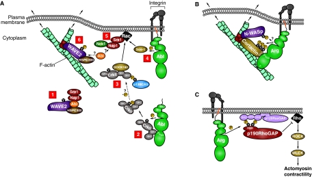
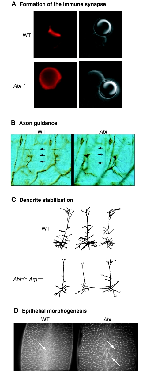
The Abl-family non-receptor tyrosine kinases are essential regulators of the cytoskeleton. They transduce diverse extracellular cues into cytoskeletal rearrangements that have dramatic effects on cell motility and morphogenesis. Recent biochemical and genetic studies have revealed several mechanisms that Abl-family kinases use to mediate these effects. Abl-family kinases stimulate actin polymerization through the activation of cortactin, hematopoietic lineage cell-specific protein (HS1), WASp- and WAVE-family proteins, and Rac1. They also attenuate cell contractility by inhibiting RhoA and altering adhesion dynamics. These pathways impinge on several physiological processes, including development and maintenance of the nervous and immune systems, and epithelial morphogenesis. Elucidating how Abl-family kinases are regulated, and where and when they coordinate cytoskeletal changes, is essential for garnering a better understanding of these complex processes.


Figures

Fig. 1.
Fig. 1.
Domain structure of Abl-family kinases. Abl and Arg each have two isoforms, the shorter non-myristoylated isoform 1a (Abl isoform I in mice) and the longer myristoylated isoform 1b (Abl isoform IV in mice). Both Abl and Arg have a `cap' region that extends from their N-terminus to their SH3 domain. C-terminal to the cap, they have sequential SH3 (red), SH2 (blue) and kinase (green) domains. Abl has four PxxP motifs (yellow stripes) and Arg has three. Abl also has nuclear localization sequences (NLS, gray stripes), a DNA-binding region (DNA, magenta) and a nuclear export sequence (NES, light-blue stripe). Additionally, Abl has G-actin (G, orange) and F-actin (F, purple)-binding domains. Arg has two F-actin-binding domains and an MT-binding domain (MT, gray). Abl and Arg isoforms 1b are 1142 and 1182 amino acids in mice, respectively. They share >90% sequence identity in their SH3, SH2 and kinase domains, 40% identity in their proline-rich regions, 45% identity in their C-terminal F-actin-binding domain, but only 21% identity in the remaining regions in the C-terminus. The related tyrosine kinase Src is myristoylated and contains SH3 and SH2 domains and a kinase domain, which share 37% and 52% identity, respectively, to the corresponding domains in Abl-family kinases. Phosphorylation of the linker and activation-loop tyrosine residues (Y in black circles) of Abl and Arg are required for full kinase activation. The linker tyrosine is between the SH2 and kinase domains (Abl Y245, Arg Y272), and the activation-loop tyrosine is in the kinase domain (Abl Y412, Arg Y439). Src also has an activation-loop tyrosine residue in its kinase domain (Src Y416). Additionally, Src has a tyrosine residue near its C-terminus that stabilizes its auto-inhibited conformation when phosphorylated (Src Y527).
Fig. 2.
Fig. 2.
Regulation of kinase activity of Abl and Arg. (A) Abl-family kinases are held in an inactive closed conformation, in which their SH3 (3, red) and SH2 (2, blue) domains fold back on their kinase domain (green). The N-terminal cap (dotted gray line) makes additional inhibitory contacts with the SH3 and SH2 domains, and allows docking of the myristoyl group (orange) in a hydrophobic pocket in the kinase domain. The C-terminal half is shown in gray. (B,C) Partial activation is achieved through interaction with receptors, such as (B) an activated integrin heterodimer (gray) or (C) cytoplasmic PxxP and/or phosphotyrosine (P in yellow circle attached to Y in black circle)-containing ligands (yellow) that open the auto-inhibited conformation. (D) Full activation is achieved by phosphorylation of the linker tyrosine (between the SH2 and kinase domains) and the activation-loop tyrosine (between the two lobes of the kinase domain) in trans by other Abl-family kinases and/or by Src-family kinases (yellow). In this example, integrin clustering brings Abl- and Src-family kinases into close proximity.
Fig. 3.
Fig. 3.
Abl-family kinases modulate cytoskeletal signaling pathways. (A) Abl modulates WAVE2-mediated actin polymerization. [1] WAVE2 (purple) is held in a trans-inhibited complex with HSPC300 (brown), Abi (orange), and Nap1 and Sra1 (red). [2] Abl (green) uses a PxxP motif to bind to a CrkII SH3 domain (gray). Abl phosphorylates CrkII, leading to an intramolecular interaction between CrkII's SH2 domain and the phosphotyrosine residue (Y221). Phosphorylated CrkII then localizes to the cell periphery. [3] At the cell periphery, CrkII is possibly dephosphorylated, allowing interaction with p130CAS (blue) and the Rac GEF DOCK180 (light brown). [4] Abl also phosphorylates and activates the Rac GEF Sos-1 (light brown). [5] Active Rac (black) interacts with Sra1, and Nck1 (dark green) interacts with Nap1, which releases the inhibitory Sra1-Nap1 sub-complex from WAVE2. It is unclear whether Abi remains bound to active WAVE2 or is released with the inhibitory sub-complex. [6] Active WAVE2 remains bound to HSPC300, and possibly Abi, can be phosphorylated by Abl and interacts with the Arp2/3 complex (red) to stimulate actin polymerization at a nascent F-actin branch (turquoise). (B) Arg modulates cortactin- and N-WASp-mediated actin polymerization. Arg (green) uses a PxxP motif to bind to the SH3 domain of cortactin (light brown) and an F-actin-binding domain (`F') to bind to F-actin (turquoise). Cortactin interacts with the Arp2/3 complex (red) and modestly stimulates actin polymerization on a nascent F-actin branch. Following integrin (gray) adhesion, active Arg phosphorylates (P in yellow circle) cortactin and possibly N-WASp (purple), leading to a greater enhancement of actin polymerization and membrane protrusion. Arg can also use its SH2 domain to interact with phosphotyrosine residues on cortactin (not shown). Cortactin and N-WASp can also bind directly to each other, as well as to the Nck1 adaptor protein (not shown). (C) Arg modulates p190RhoGAP-mediated inhibition of RhoA. Following integrin (gray) adhesion, Arg phosphorylates the RasGAP-binding-region (RBR) of p190RhoGAP (p190; red), which then interacts with the SH2 domains of p120RasGAP (p120; light purple). p120RasGAP has phospholipid-binding domains (PH and C2), which allow the p190-p120 complex to localize to the membrane. Once in proximity to active Rho (black), p190 inhibits Rho by stimulating Rho's weak intrinsic GTPase activity. Active Rho normally activates ROCK (light brown), which, in turn, activates MLCK (light brown) to phosphorylate myosin, leading to increased actomyosin contractility. Therefore, p190 acts as a brake on actomyosin contractility.
Fig. 4.
Fig. 4.
Physiological roles for Abl-family kinases. (A) Abl is required for proper F-actin assembly during immune-synapse formation. Wild-type (WT, top) or Abl–/– (bottom) T cells were incubated with latex beads (right panels) coated with anti-T-cell-receptor antibody, and were then stained with phalloidin to reveal the F-actin cytoskeleton (left panels). Wild-type T cells form an immune-synapse-like concentration of F-actin at the bead contact site. The F-actin cytoskeleton remains diffusely localized in Abl–/– T cells. Figure courtesy of Ann Huang and Janis Burkhardt (University of Pennsylvania, Philadelphia, PA). (B) dAbl is required for proper axon guidance. In abdominal segments of wild-type (WT, left) Drosophila embryos, intersegmental neuron group b (ISNb) axons make normal neuromuscular junctions with longitudinal muscles, including muscle 12 (arrows). ISNb growth cones stop short and fail to contact muscle 12 in an Abl mutant (right). Adapted with permission from figure 3 in Wills et al. (Wills et al., 1999b). Figure courtesy of David Van Vactor (Harvard Medical School, Boston, MA). (C) Abl and Arg are required for dendrite stabilization. Camera lucida representations of wild-type (WT, top) or Abl–/– Arg–/– (bottom) cortical-layer-5 pyramidal neurons. Atrophy of dendrite arbors in Abl–/– Arg–/– neurons results in smaller dendrite arbors. (D) dAbl is essential for normal epithelial morphogenesis. Wild-type Drosophila embryos (WT, left) exhibit uniform apical constriction at the ventral furrow during gastrulation (arrow). Cell morphology is visualized using a moesin-GFP that binds to F-actin. An Ablmz mutant depleted for both maternal and zygotic Abl (right) exhibits non-uniform constriction. Figure courtesy of Don Fox and Mark Peifer (University of North Carolina, Chapel Hill, NC).

Comment in

  • ABL at the leading edge.
    Krause M. Krause M. Nat Rev Mol Cell Biol. 2011 Jan;12(1):8. doi: 10.1038/nrm3026. Epub 2010 Nov 24. Nat Rev Mol Cell Biol. 2011. PMID: 21102623 No abstract available.

References

    1. Abassi, Y. A. and Vuori, K. (2002). Tyrosine 221 in Crk regulates adhesion-dependent membrane localization of Crk and Rac and activation of Rac signaling. EMBO J. 21, 4571-4582. - PMC - PubMed
    1. Antoku, S., Saksela, K., Rivera, G. M. and Mayer, B. J. (2008). A crucial role in cell spreading for the interaction of Abl PxxP motifs with Crk and Nck adaptors. J. Cell Sci. 121, 3071-3082. - PMC - PubMed
    1. Arias-Salgado, E. G., Lizano, S., Sarkar, S., Brugge, J. S., Ginsberg, M. H. and Shattil, S. J. (2003). Src kinase activation by direct interaction with the integrin beta cytoplasmic domain. Proc. Natl. Acad. Sci. USA 100, 13298-13302. - PMC - PubMed
    1. Arthur, W. T., Noren, N. K. and Burridge, K. (2002). Regulation of Rho family GTPases by cell-cell and cell-matrix adhesion. Biol. Res. 35, 239-246. - PubMed
    1. Backert, S., Feller, S. M. and Wessler, S. (2008). Emerging roles of Abl family tyrosine kinases in microbial pathogenesis. Trends Biochem. Sci. 33, 80-90. - PubMed

Publication types

Substances