. 2009 Sep 16;29(37):11674-85.
doi: 10.1523/JNEUROSCI.1542-09.2009.

Axonal targeting of Trk receptors via transcytosis regulates sensitivity to neurotrophin responses

Affiliations

Axonal targeting of Trk receptors via transcytosis regulates sensitivity to neurotrophin responses

Maria Ascaño et al. J Neurosci. .

Abstract

Axonal targeting of trophic receptors is critical for neuronal responses to extracellular developmental cues, yet the underlying trafficking mechanisms remain unclear. Here, we report that tropomyosin-related kinase (Trk) receptors for target-derived neurotrophins are anterogradely trafficked to axons via transcytosis in sympathetic neurons. Using compartmentalized cultures, we show that mature receptors on neuronal soma surfaces are endocytosed and remobilized via Rab11-positive recycling endosomes into axons. Inhibition of dynamin-dependent endocytosis disrupted anterograde transport and localization of TrkA receptors in axons. Anterograde TrkA delivery and exocytosis into axon growth cones is enhanced by nerve growth factor (NGF), acting locally on distal axons. Perturbing endocytic recycling attenuated NGF-dependent signaling and axon growth while enhancing recycling conferred increased neuronal sensitivity to NGF. Our results reveal regulated transcytosis as an unexpected mode of Trk trafficking that serves to rapidly mobilize ready-synthesized receptors to growth cones, thus providing a positive feedback mechanism by which limiting concentrations of target-derived neurotrophins enhance neuronal sensitivity.


Figures

Figure 1.
Figure 1.
Trk receptors undergo constitutive endocytosis and recycling in sympathetic neurons. A, Constitutively internalized TrkA receptors are sequestered intracellularly during blocking recycling with monensin (10 μm). Membrane proteins were subjected to cell-surface biotinylation. Internalized TrkA receptors were detected by surface stripping of biotin, neutravidin precipitation, and TrkA Western blotting. B, Densitometric quantification of the results shown in A. Results are the mean ± SEM from seven independent experiments. *p < 0.05, t test. C, Monensin-dependent intracellular accumulation of surface TrkA receptors is attenuated in neurons expressing Dyn–K44A. Cell-surface biotinylation assay was performed in neurons infected with adenoviral vectors expressing GFP and Dyn–K44A, in the presence of monensin or NGF. Normalization for protein amounts in A and C was performed by immunoblotting neuronal lysates for p85 subunit of PI3K. D, Live antibody feeding assays in neurons expressing FLAG–TrkB:A chimeric receptors show subcellular localization of receptors under the conditions indicated. Scale bar, 5 μm. E, Quantification of internal accumulation of chimeric receptors under the various conditions indicated in D, by assessing the proportion of colocalization of FLAG immunofluorescence with that of GFP (data not shown), which is coexpressed in infected neurons and is cytoplasmic. Values have been corrected by subtracting any colocalization observed at 4°C. Results are the mean ± SEM from five independent experiments. *p < 0.05 relative to No Ligand condition, ANOVA, followed by a Tukey's post hoc test. F, Schematic of a live-cell ratiometric assay to measure receptor internalization and recycling. G, Spatial analyses of constitutive Trk trafficking in neurons. Representative images of surface, constitutive internalized, and recycled chimeric receptors in cell bodies (top row) and axons (bottom row), as assessed by the live-cell ratiometric assay performed independently in cell body and axon compartments in compartmentalized neuronal cultures. Scale bars: Cell Body, 5 μm; Axon, 20 μm. Graphs represent quantification of Trk internalization (H) and recycling (I) from seven independent experiments. Values are means ± SEM; **p = 0.02, t test.
Figure 2.
Figure 2.
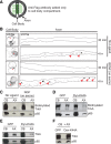
Trk receptors are transcytosed from neuronal soma surfaces to axons. A, Schematic of antibody feeding assay in compartmentalized cultures expressing FLAG–TrkB:A to follow transcytosis of Trk receptors originating from neuronal soma surfaces. B, Representative images of cell bodies and axonal projections fixed and stained for anti-FLAG antibody. Anti-FLAG-labeled TrkB:A receptors are evident in cell bodies at 30 min and 4 h (small arrowheads) and in axons only in the 4 h incubations (large arrowheads). Scale bars, 5 μm. C, NGF treatment of axons (100 ng/ml, 4 h) enhances levels of biotinylated TrkA receptors in axons. Lysates from cell body (CB) and axon (AX) compartments in compartmentalized cultures were precipitated with neutravidin and immunoblotted with TrkA. Normalization for protein amounts was performed by immunoblotting for p85 subunit of PI3K. D, Dominant-negative dynamin (Dyn–K44A) inhibits TrkA transcytosis. Cell-surface biotinylation assay was performed in compartmentalized cultures expressing GFP or Dyn–K44A and treated with NGF on axons (100 ng/ml, 4 h). E, F, Dyn–K44A leads to a decrease in axonal, but not total, TrkA levels. Western blot analysis of TrkA levels in compartmentalized cultures (E) shows that expression of Dyn–K44A leads to a significant decrease in axonal TrkA levels. Dyn–K44A does not affect total TrkA levels in neurons (F).
Figure 3.
Figure 3.
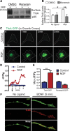
Rab11a mediates Trk transcytosis in sympathetic neurons. A, FLAG–TrkB:A receptors undergoing transcytosis colocalize with Rab11a in axons (arrowheads) as detected by immunostaining for FLAG antibody and endogenous Rab11a. Antibody-feeding assay was performed on compartmentalized cultures expressing FLAG–TrkB:A in the presence of BDNF (100 ng/ml, 4 h) on axons. Boxed region in the middle is shown at higher magnification on the right. B, FLAG–TrkB:A receptors undergoing transcytosis colocalize with Alexa-546-labeled Tf (Alexa-546–Tf) in axons (arrowheads) as detected by immunostaining for FLAG antibody and Alexa-546–Tf fluorescence. Cell body compartments of compartmentalized cultures expressing FLAG–TrkB:A were fed with FLAG antibodies and Alexa-546–Tf under live conditions. After 4 h of labeling, neurons were fixed, permeabilized, and labeled with anti-mouse fluorescent secondary antibody to visualize FLAG-antibody-bound receptors. Boxed region in the middle is shown at higher magnification on the right. C, Individual frames from time-lapse microscopy of neurons expressing TrkA–GFP and mcherry–Rab11a, with corresponding kymograph (bottom). Anterogradely and retrogradely moving double-positive carriers are indicated by white arrowhead and white arrow, respectively, and purple arrowhead indicates a retrogradely moving structure positive for TrkA–GFP alone. Scale bars: A–C, 5 μm. D, Histogram showing speed distribution of mobile (>0.6 μm/s) carriers double positive for TrkA–GFP and mcherry–Rab11a. Quantification of 3289 single movements [defined as the distance traversed by a carrier between two consecutive frames (2 s)] of 253 double-positive structures from a total of four independent experiments. E, Rab11a activity is required for TrkA transcytosis, as assessed by the biotinylation assay in compartmentalized cultures expressing GFP or Rab11a–S25N. AX, Axon; CB, cell body.
Figure 4.
Figure 4.
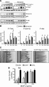
NGF stimulation promotes rapid exocytosis of TrkA from recycling endosomes. A, TrkA is rapidly recruited to the cell surface during NGF stimulation. Levels of surface TrkA were assessed using cell-surface biotinylation in neuronal cultures briefly stimulated with a 5 min pulse of NGF (100 ng/ml) in the presence or absence of the recycling blocker monensin. Normalization for protein amounts was performed by immunoblotting neuronal lysates for p85 subunit of PI3K. B, Densitometric quantification of the results shown in A. Results are quantified relative to the NGF (100 ng/ml) condition, from eight independent experiments. *p < 0.05 and **p < 0.01 as determined by one-way ANOVA, followed by a Dunnett's multiple comparison test. C, TIRF analyses of growth cones from neurons expressing TrkA–GFP. Dashed lines outline the periphery of growth cones. Scale bar, 5 μm. D, Time course of TrkA–GFP fluorescence in the two growth cones depicted in C. Arrow indicates time of NGF addition. E, Group data showing maximum change in fluorescence (1–5 min) from growth cones of neurons expressing TrkA–GFP (n = 15) or farnesylated EGFP. Values are means ± SEM from four independent experiments. **p = 0.003, t test. F, Rapid BDNF-mediated surface delivery of FLAG–TrkB:A receptors originating from the plasma membrane. FLAG–TrkB:A receptors were live labeled with FLAG antibodies and allowed to internalize, surface antibodies were stripped off, and neurons were stimulated with BDNF for 5 min in the presence of a fluorescent secondary antibody.
Figure 5.
Figure 5.
NGF-induced TrkA exocytosis modulates neuronal sensitivity to NGF-dependent signaling and axon growth. A, Blocking endocytic recycling attenuates NGF-dependent signaling. Neuronal cultures were briefly stimulated (5 min) with indicated concentrations of NGF in the presence or absence of the recycling blocker monensin (10 μm). Lysates were immunoblotted to detect P-TrkA, P-Erk1/2, and P-Akt. Normalization for protein amounts was performed by stripping the immunoblots and reprobing for p85. B, Densitometric quantification of the results shown in A. Results are quantified relative to the NGF (1000 ng/ml) condition, from seven independent experiments. *p < 0.05 as determined by two-way ANOVA, followed by Bonferroni's post hoc test. C, D, Rab11a activity regulates NGF-dependent axon growth. Compartmentalized cultures were infected with adenoviruses expressing GFP or Rab11 constructs, EGFP–Rab11a–S25N (dominant-negative) and EGFP–Rab11a–Q70L (constitutively active), and maintained with NGF added solely to the axonal compartments. Rate of axon extension (micrometers per day) was assessed for 72 h. Panels in C are representative images of axons immunostained with anti-β-III tubulin. Separation between cell body (CB) and axon (AX) compartments is indicated by dashed line. D, Graph shows quantification of axon growth. Values are means ± SEM from seven independent experiments. *p < 0.05 and **p < 0.01, two-way ANOVA analysis with Bonferroni's post hoc test.
Figure 6.
Figure 6.
Model for TrkA transcytosis in sympathetic neurons. Newly synthesized TrkA receptors are first inserted into the neuronal soma surfaces, where they undergo constitutive endocytosis and subsequent axonal targeting via Rab11-positive recycling endosomes (blue arrows). In the absence of ligand, more receptors are internalized in neuronal soma compared with axons, promoting a net flow toward axons. NGF signaling initiated at axon terminals triggers a transient increase in surface TrkA levels through local exocytosis from intracellular receptor pools, followed by endocytosis and retrograde transport of activated receptors in nerve terminals (gray arrows). Retrograde NGF signaling enhances transcytosis leading to the anterograde delivery of additional TrkA receptors. The contribution of the secretory pathway to axonal targeting of Trk receptors remains to be determined.

References

    1. Altschuler Y, Hodson C, Milgram SL. The apical compartment: trafficking pathways, regulators and scaffolding proteins. Curr Opin Cell Biol. 2003;15:423–429. - PubMed
    1. Anderson E, Maday S, Sfakianos J, Hull M, Winckler B, Sheff D, Fölsch H, Mellman I. Transcytosis of NgCAM in epithelial cells reflects differential signal recognition on the endocytic and secretory pathways. J Cell Biol. 2005;170:595–605. - PMC - PubMed
    1. Atwal JK, Massie B, Miller FD, Kaplan DR. The TrkB-Shc site signals neuronal survival and local axon growth via MEK and P13-kinase. Neuron. 2000;27:265–277. - PubMed
    1. Bastaki M, Braiterman LT, Johns DC, Chen YH, Hubbard AL. Absence of direct delivery for single transmembrane apical proteins or their “secretory” forms in polarized hepatic cells. Mol Biol Cell. 2002;13:225–237. - PMC - PubMed
    1. Basu SK, Goldstein JL, Anderson RG, Brown MS. Monensin interrupts the recycling of low density lipoprotein receptors in human fibroblasts. Cell. 1981;24:493–502. - PubMed

Publication types

MeSH terms