. 2009 Dec;76(6):1341-8.
doi: 10.1124/mol.109.058578. Epub 2009 Sep 16.

Quantification of Gi-mediated inhibition of adenylyl cyclase activity reveals that UDP is a potent agonist of the human P2Y14 receptor

Affiliations

Quantification of Gi-mediated inhibition of adenylyl cyclase activity reveals that UDP is a potent agonist of the human P2Y14 receptor

Rhonda L Carter et al. Mol Pharmacol. 2009 Dec.

Abstract

The P2Y14 receptor was initially identified as a G protein-coupled receptor activated by UDP-glucose and other nucleotide sugars. We have developed several cell lines that stably express the human P2Y14 receptor, allowing facile examination of its coupling to native Gi family G proteins and their associated downstream signaling pathways (J Pharmacol Exp Ther 330:162-168, 2009). In the current study, we examined P2Y14 receptor-dependent inhibition of cyclic AMP accumulation in human embryonic kidney (HEK) 293, C6 glioma, and Chinese hamster ovary (CHO) cells stably expressing this receptor. Not only was the human P2Y14 receptor activated by UDP-glucose, but it also was activated by UDP. The apparent efficacies of UDP and UDP-glucose were similar, and the EC50 values (74, 33, and 29 nM) for UDP-dependent activation of the P2Y14 receptor in HEK293, CHO, and C6 glioma cells, respectively, were similar to the EC50 values (323, 132, and 72 nM) observed for UDP-glucose. UDP and UDP-glucose also stimulated extracellular signal-regulated kinase (ERK) 1/2 phosphorylation in P2Y14 receptor-expressing HEK293 cells but not in wild-type HEK293 cells. A series of analogs of UDP were potent P2Y14 receptor agonists, but the naturally occurring nucleoside diphosphates, CDP, GDP, and ADP exhibited agonist potencies over 100-fold less than that observed with UDP. Two UDP analogs were identified that selectively activate the P2Y14 receptor over the UDP-activated P2Y6 receptor, and these molecules stimulated phosphorylation of ERK1/2 in differentiated human HL-60 promyeloleukemia cells, which natively express the P2Y14 receptor but had no effect in wild-type HL-60 cells, which do not express the receptor. We conclude that UDP is an important cognate agonist of the human P2Y14 receptor.


Figures

Fig. 1.
Fig. 1.
UDP-glucose and UDP promote pertussis toxin-sensitive inhibition of cyclic AMP accumulation in P2Y14-HEK293 cells. A, the capacity of 100 μM UDP or 100 μM UDP-glucose (UDPG) to inhibit forskolin (Fsk; 30 μM)-stimulated cyclic AMP accumulation was quantified in wild-type HEK293 cells as described under Materials and Methods. The data are mean ± S.E.M. from triplicate determinations and are representative of three separate experiments. B, concentration-dependent inhibition of forskolin-stimulated cyclic AMP accumulation by UDP-glucose and UDP. P2Y14-HEK293 cells were incubated with 30 μM forskolin alone or 30 μM forskolin plus the indicated concentrations of UDP-glucose (■) or UDP (□). The data are presented as mean ± S.E.M. of triplicate determinations and are representative of data from six experiments. C, blockade of the P2Y14-R-dependent effects of UDP-glucose and UDP by pertussis toxin (PT). P2Y14-HEK293 cells were incubated overnight with vehicle or 100 ng/ml pertussis toxin, and cyclic AMP accumulation was subsequently measured in the presence of 30 μM forskolin alone or with 30 μM forskolin plus 10 μM UDP-glucose or 10 μM UDP. The data are presented as mean ± S.E.M. of results from three separate experiments.
Fig. 2.
Fig. 2.
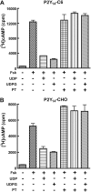
P2Y14-R-dependent activities of UDP-glucose and UDP in P2Y14-C6 and P2Y14-CHO cells. A, the capacity of 100 μM UDP or 100 μM UDP-glucose (UDPG) to inhibit forskolin (30 μM)-stimulated cyclic AMP accumulation was quantified in wild-type C6 rat glioma cells as described under Materials and Methods. The data are mean ± S.E.M. from triplicate determinations and are representative of results from three separate experiments. B, concentration-dependent inhibition of forskolin-stimulated cyclic AMP accumulation by UDP-glucose and UDP in P2Y14-C6 cells. P2Y14-C6 cells were incubated in the presence of 30 μM forskolin alone or with 30 μM forskolin plus the indicated concentrations of UDP-glucose (■) or UDP (□). The data are presented as mean ± S.E.M. of triplicate determinations and are representative of data from six separate experiments. C, concentration-dependent inhibition of forskolin-stimulated cyclic AMP accumulation by UDP-glucose and UDP in P2Y14-CHO cells. P2Y14-CHO cells were incubated in the presence of 30 μM forskolin alone or with 30 μM forskolin plus the indicated concentrations of UDP-glucose (■) or UDP (□). The data are presented as mean ± S.E.M. of triplicate determinations and are representative of data from six separate experiments.
Fig. 3.
Fig. 3.
Pertussis toxin sensitivity of the effects of UDP-glucose and UDP in P2Y14-C6 cells and P2Y14-CHO cells. A, blockade of the P2Y14-R-dependent effects of UDP-glucose and UDP by pertussis toxin in P2Y14-C6 cells. P2Y14-C6 cells were incubated overnight with vehicle or 100 ng/ml pertussis toxin, and cyclic AMP accumulation was subsequently measured in the presence of 30 μM forskolin alone or with 30 μM forskolin plus 10 μM UDP-glucose or 10 μM UDP. The data are presented as mean ± S.E.M. of results from three separate experiments. B, blockade of the P2Y14-R-dependent effects of UDP-glucose and UDP by pertussis toxin in P2Y14-CHO cells. P2Y14-CHO cells were incubated overnight with vehicle or 100 ng/ml pertussis toxin, and cyclic AMP accumulation was subsequently measured in the presence of 30 μM forskolin alone or with 30 μM forskolin plus 10 μM UDP-glucose or 10 μM UDP. The data are presented as mean ± S.E.M. of results from three separate experiments.
Fig. 4.
Fig. 4.
Agonist activities of nucleoside diphosphate molecules in P2Y14-HEK293 cells. P2Y14-HEK293 cells were incubated in the presence of 30 μM forskolin alone or with 30 μM forskolin plus the indicated concentrations of UDP (□), CDP (▿), GDP (◆), or ADP (∗). The data are presented as mean ± S.E.M. of results pooled from three separate experiments.
Fig. 5.
Fig. 5.
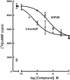
Agonist activities of 2-thio-UDP and UDPβS in P2Y14-HEK293 cells. P2Y14-HEK293 cells were incubated in the absence (○) or presence of 30 μM forskolin alone or with 30 μM forskolin plus the indicated concentrations of 2-thio-UDP (▾) or UDPβS (▵). The data are presented as mean ± S.E.M. of triplicate determinations and are representative of data from three separate experiments.
Fig. 6.
Fig. 6.
P2Y14-R-dependent activation of MAP kinase signaling by UDP. Empty vector or P2Y14-HEK293 cells were serum-starved for 18 h before incubation with vehicle, 10 μM UDP, or 10 μM UDP-glucose for 15 min. Cell lysates were subjected to SDS-polyacrylamide gel electrophoresis, the samples transferred to nitrocellulose membranes, and the membranes probed with antibodies for phospho-ERK1/2 and total ERK1/2 as described under Materials and Methods. The results shown are representative of data from three individual experiments.
Fig. 7.
Fig. 7.
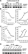
Activation of a P2Y14-R-dependent MAP kinase signaling response in differentiated human HL-60 promyeloleukemia cells. A, phosphorylation of ERK1/2 (phospho-ERK1/2) was analyzed in wild-type HL-60 cells after incubation for the indicated times with 10 μM UDP or 10 μM UDP-glucose (UDPG) as described under Materials and Methods. Levels of total ERK1/2 also are presented. B, concentration effect curves for MRS2802 for activation of P2Y14-R in P2Y14-HEK293 cells and activation of P2Y6-R in 1321N1 human astrocytoma cells stably expressing the human P2Y6-R were generated as described under Materials and Methods. C, concentration effect curves for MRS2907 for activation of P2Y14-R in P2Y14-HEK293 cells and activation of P2Y6-R in 1321N1 human astrocytoma cells stably expressing the human P2Y6-R were generated as described under Materials and Methods. D, phosphorylation of ERK1/2 (phospho-ERK1/2) was quantified in undifferentiated (−DMSO) or differentiated (+DMSO) HL-60 cells after incubation for 30 min with 10 μM UDP-glucose (UDPG), 1 μM MRS2907, or 10 μM MRS2802 as described under Materials and Methods. Levels of total ERK1/2 also are presented.

References

    1. Abbracchio MP, Burnstock G, Boeynaems JM, Barnard EA, Boyer JL, Kennedy C, Knight GE, Fumagalli M, Gachet C, Jacobson KA, Weisman GA. (2006) International Union of Pharmacology LVIII: update on the P2Y G protein-coupled nucleotide receptors: from molecular mechanisms and pathophysiology to therapy. Pharmacol Rev 58:281–341 - PMC - PubMed
    1. Alvarado-Castillo C, Harden TK, Boyer JL. (2005) Regulation of P2Y1 receptor-mediated signaling by the ectonucleoside triphosphate diphosphohydrolase isozymes NTPDase1 and NTPDase2. Mol Pharmacol 67:114–122 - PubMed
    1. Ault AD, Broach JR. (2006) Creation of GPCR-based chemical sensors by directed evolution in yeast. Protein Eng Des Sel 19:1–8 - PubMed
    1. Besada P, Shin DH, Costanzi S, Ko H, Mathé C, Gagneron J, Gosselin G, Maddileti S, Harden TK, Jacobson KA. (2006) Structure-activity relationships of uridine 5′-diphosphate analogues at the human P2Y6 receptor. J Med Chem 49:5532–5543 - PMC - PubMed
    1. Burnstock G. (2007) Physiology and pathophysiology of purinergic neurotransmission. Physiol Rev 87:659–797 - PubMed

Publication types

LinkOut - more resources