. 2009 Nov 12;114(20):4451-9.
doi: 10.1182/blood-2009-07-233346. Epub 2009 Sep 16.

Stage-specific Arf tumor suppression in Notch1-induced T-cell acute lymphoblastic leukemia

Affiliations

Stage-specific Arf tumor suppression in Notch1-induced T-cell acute lymphoblastic leukemia

Emmanuel J Volanakis et al. Blood. .

Abstract

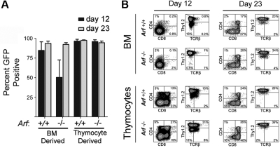
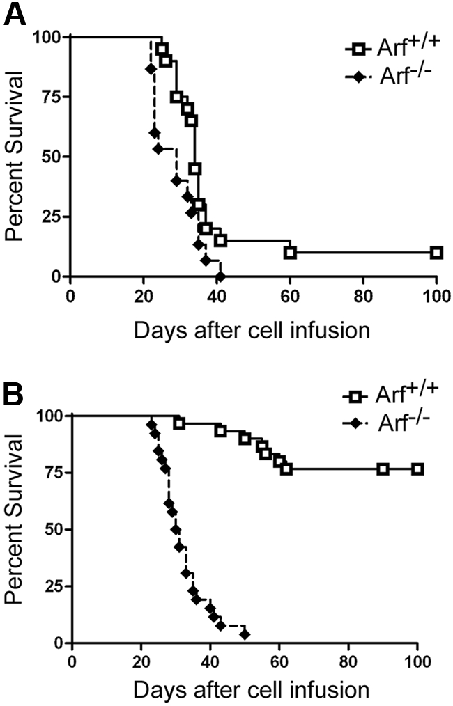
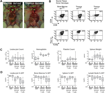
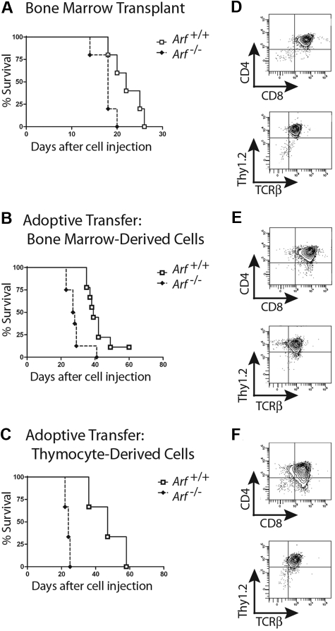
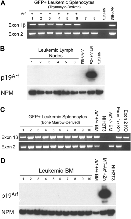
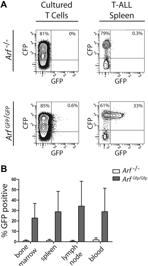
Frequent hallmarks of T-cell acute lymphoblastic leukemia (T-ALL) include aberrant NOTCH signaling and deletion of the CDKN2A locus, which contains 2 closely linked tumor suppressor genes (INK4A and ARF). When bone marrow cells or thymocytes transduced with a vector encoding the constitutively activated intracellular domain of Notch1 (ICN1) are expanded ex vivo under conditions that support T-cell development, cultured progenitors rapidly induce CD4+/CD8+ T-ALLs after infusion into healthy syngeneic mice. Under these conditions, enforced ICN1 expression also drives formation of T-ALLs in unconditioned CD-1 nude mice, bypassing any requirements for thymic maturation. Retention of Arf had relatively modest activity in suppressing the formation of T-ALLs arising from bone marrow-derived ICN1+ progenitors in which the locus is epigenetically silenced, and all resulting Arf (+/+) tumors failed to express the p19(Arf) protein. In striking contrast, retention of Arf in thymocyte-derived ICN1+ donor cells significantly delayed disease onset and suppressed the penetrance of T-ALL. Use of cultured thymocyte-derived donor cells expressing a functionally null Arf-GFP knock-in allele confirmed that ICN1 signaling can induce Arf expression in vivo. Arf activation by ICN1 in T cells thereby provides stage-specific tumor suppression but also a strong selective pressure for deletion of the locus in T-ALL.


Figures

Figure 1
Figure 1
Emergence of GFP+ cells and their immunophenotypes on day 12 and day 23 of culture. (A) Cells derived from 5-FU–conditioned bone marrow (BM) or from thymocytes explanted from mice of the indicated Arf genotypes (labeled at bottom) were transduced with a vector encoding ICN1 and GFP in cis and cultured on OP9 stroma with cytokine support. Cultured cells expanded exponentially and underwent 25 to 30 population doublings over a period of 36 days (supplemental Figure 1). The percentage of GFP+ cells in the cultures was determined at day 12 and day 23. The results of 3 experiments are shown, and error bars indicate SDs. (B) After 12 and 23 days of culture (indicated at the top of the panels) the percentages of GFP+ cells that expressed CD4 and CD8 or Thy1.2 and TCRβ were determined using an automated cell sorter. The percentages of cells expressing each marker are indicated in at least 3 of the 4 quadrants of each panel. The origin of donor cells and their Arf genotypes are indicated to the left.
Figure 2
Figure 2
Survival curves of recipient mice developing T-ALL. Survival curves are shown for cohorts of healthy nonirradiated C57BL/6 mice injected with cultured (day-12) ICN1+ cells derived from the bone marrow of 5-FU–conditioned mice (A) or unfractionated thymocytes (B) of the indicated Arf genotypes. (A) Twenty recipients of Arf +/+ and 15 recipients of Arf −/− bone marrow–derived cells in 2 experiments had a median survival of 34 days and 29 days, respectively. The differences in latency are statistically significant (P = .02 by log-rank test). (B) In 3 experiments, all but 7 of 30 recipients of Arf +/+ thymus-derived cells did not develop T-ALL, whereas 25 of 26 recipients of Arf −/− cells died of disease with a median survival of 30.5 days. Differences in latency are highly significant (P < .001 by log-rank test).
Figure 3
Figure 3
T-ALLs arising from cultured ICN1+ cells have similar disease phenotypes whether derived from bone marrow or thymocytes. (A) Moribund mice were killed, dissected, and photographed. Images were captured with a Nikon Coolpix 5200 camera (Nikon) in ambient room light and through a Tiffen 15 deep yellow filter (Tiffen) under fluorescent illumination provided by an Illumatool light source (Light Tools Research). The images were merged using Photoshop 7.0 software (Adobe Systems). Recipient animals developed T-ALL with hepatosplenomegaly and extensive invasion of lymph nodes with GFP-marked leukemic cells. Representative recipients of marrow-derived cells (left panel) and of thymocyte-derived cells (right panel) are depicted. (B) Like irradiated animals that received bone marrow transplants of ICN1-transduced, but uncultured, donor cells, the T-ALLs arising in nonirradiated mice largely coexpressed CD4 and CD8, though some T-ALLs had a CD8 single-positive subpopulation, as well. No consistent variations in representative immunophenotypes of leukemic cells taken from different tissues or derived from Arf +/+ or Arf −/− donors were seen. The remaining panels (C-D) compare clinical parameters of T-ALLs arising in irradiated animals undergoing conventional bone marrow transplantations (BMTs) with those of recipient mice that received cultured day-12 ICN1+ bone marrow–derived or thymus-derived cells. Clinical hematologic measurements including a comparison of total white blood cell counts, hemoglobin concentration, platelet count, and spleen weights are shown in panel C, whereas the percentages of GFP-marked cells in various tissues are shown in panel D. The broad horizontal bars indicate median values, the gray bars delineate the upper and lower quartiles, and the whiskers indicate the overall range of readings.
Figure 4
Figure 4
Athymic nude mice develop CD4+/CD8+ T-ALLs when infused with ICN1-expressing cells. Survival curves are shown for cohorts of irradiated nude mice infused with noncultured ICN1+ bone marrow–derived cells (A; 5 mice/group; P = .03 by log-rank test), nonirradiated nude mice infused with ICN1+ bone marrow–derived cells after 12 days of culture (B; 9 Arf +/+, 8 Arf −/− recipient mice, P = .002), and nonirradiated nude mice infused with cultured ICN1+ thymus-derived cells (C; 3 mice/group, P = .02). Representative immunophenotypes of T-ALLs arising in each experiment are depicted in panels to the right of the corresponding survival curves (D-F). Although all leukemias had a dominant CD4+/CD8+ population, several also had CD8 single-positive subpopulations.
Figure 5
Figure 5
T-ALLs arising from cultured ICN1+Arf+/+ donor cells retain the gene but do not express p19Arf protein. (A) Leukemic GFP+ splenocytes from 8 mice that had received cultured (day-12) thymocyte-derived donor cells of the indicated Arf genotypes (top) were purified by FACS. Genomic DNA prepared from these cells was used as a template for PCR performed with primers specific for exon-1β and exon-2 of the Ink4a-Arf locus. Exon-1β encodes the unique N-terminus of p19Arf, whereas exon-2 encodes C-terminal portions of p19Arf and p16Ink4a from alternative reading frames. DNAs extracted from unmanipulated Arf +/+ bone marrow cells and from mouse NIH-3T3 cells that had deleted the Ink4a-Arf locus during the process of immortalization were used as the positive and negative controls, respectively. The fact that no exon-1β signal was revealed in samples 7 and 8 generated from Arf −/− donor cells indicates that the purified leukemic cells were uncontaminated by normal Arf +/+ cells. (B) Although all genotyped leukemias retained the Arf locus, immunoblotting of electrophoretically separated proteins extracted from robustly GFP-positive lymph nodes taken from the same recipients of Arf +/+ donor cells failed to reveal p19Arf expression. Arf expression is also silenced in normal bone marrow (BM) cells. Cells engineered to express Arf conditionally under the control of a metallothionein promoter (MT-Arf cells) were induced with zinc. The immunoblot was developed with a highly sensitive monoclonal antibody generated to mouse p19Arf (Bertwistle et al), and the film was purposely overexposed in an attempt to reveal p19Arf expression. Nucleophosmin (NPM), an abundant nucleolar protein, was used as the loading control. (C) PCR analysis as in panel A. Samples 1 to 4 were taken from irradiated recipients that had received a transplant of uncultured ICN1-expressing bone marrow from 5-FU–conditioned donors. Samples 5 to 10 were taken from nonirradiated recipients that had received 5-FU–conditioned bone marrow transduced with ICN1 and cultured on OP9 stroma before their adoptive transfer. Control PCR reactions were performed using DNAs extracted from unmanipulated Arf +/+ bone marrow cells, NIH-3T3 cells lacking the Ink4a-Arf locus, bone marrow cells from Arf −/− mice in which exon-1β is disrupted, bone marrow cells from p16Ink4a-null mice in which the unique Ink4a exon-1α was disrupted, and from mice in which Ink4a-Arf exon-2 was disrupted. (D) Immunoblotting for p19Arf was performed on bone marrow taken from the mice analyzed in panel C.
Figure 6
Figure 6
Activation of the Arf promoter during T-ALL development. Healthy syngeneic C57BL/6 mice received donor T cells derived from either control Arf −/− or “knock-in” Arf Gfp/Gfp (also functionally null) thymocytes infected with an ICN1-CFP vector. (A) Cells from day-12 cultured thymocytes (left panels) or from spleens of moribund mice (right panels) were studied by FACS for coexpression of vector-coded CFP (ordinate) and cellular Arf-encoded GFP (abscissa). (B) Hematopoietic tissues from 17 such mice were similarly analyzed and contained a significant proportion of CFP-marked cells that coexpressed GFP (ordinate). Error bars indicate the SD from the mean.

References

    1. Pui C-H, Robison LL, Look AT. Acute lymphoblastic leukemia. Lancet. 2008;371(9617):1030–1043. - PubMed
    1. Weng AP, Ferrando AA, Lee W, et al. Activating mutations of NOTCH1 in human T cell acute lymphoblastic leukemia. Science. 2004;306(5694):269–271. - PubMed
    1. Irvine KD, Rauskolb C. Boundaries in development: formation and function. Annu Rev Cell Dev Biol. 2001;17:189–214. - PubMed
    1. Aster JC, Pear WS, Blacklow SC. Notch signaling in leukemia. Annu Rev Pathol. 2008;3:587–613. - PMC - PubMed
    1. Grabher C, von Boehmer H, Look AT. Notch 1 activation in the molecular pathogenesis of T-cell acute lymphoblastic leukemia. Nat Rev Cancer. 2006;6(5):347–359. - PubMed

Publication types

MeSH terms

Substances