. 2009 Nov 26;114(23):4784-91.
doi: 10.1182/blood-2009-07-230482. Epub 2009 Sep 16.

Generation of Epstein-Barr virus-specific cytotoxic T lymphocytes resistant to the immunosuppressive drug tacrolimus (FK506)

Affiliations

Generation of Epstein-Barr virus-specific cytotoxic T lymphocytes resistant to the immunosuppressive drug tacrolimus (FK506)

Biagio De Angelis et al. Blood. .

Abstract

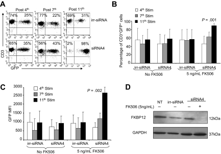
Adoptive transfer of autologous Epstein-Barr virus-specific cytotoxic T lymphocytes (EBV-CTLs) to solid organ transplant (SOT) recipients has been shown safe and effective for the treatment of EBV-associated posttransplantation lymphoproliferative disorders (PTLDs). SOT recipients, however, require the continuous administration of immunosuppressive drugs to prevent graft rejection, and these agents may significantly limit the long-term persistence of transferred EBV-CTLs, precluding their use as prophylaxis. Tacrolimus (FK506) is one of the most widely used immunosuppressive agents in SOT recipients, and its immunosuppressive effects are largely dependent on its interaction with the 12-kDa FK506-binding protein (FKBP12). We have knocked down the expression of FKBP12 in EBV-CTLs using a specific small interfering RNA (siRNA) stably expressed from a retroviral vector and found that FKBP12-silenced EBV-CTLs are FK506 resistant. These cells continue to expand in the presence of the drug without measurable impairment of their antigen specificity or cytotoxic activity. We confirmed their FK506 resistance and anti-PTLD activity in vivo using a xenogenic mouse model, suggesting that the proposed strategy may be of value to enhance EBV-specific immune surveillance in patients at high risk of PTLD after transplantation.


Figures

Figure 1
Figure 1
Identification of a siRNA vector that stably knocks down FKBP12. (A) GFP MFI of 293T cells transfected with eGFP-hFKBP12 (+) or cotransfected with eGFP-hFKBP12 and each of the 17 predicted siRNA sequences targeting the human FKBP12 mRNA or an irrelevant siRNA (irr-siRNA). siRNA4 resulted in > 85% reduction of the GFP MFI. (B) GFP was expressed by 60% of 293T cells transfected with the reporter plasmid eGFP-hFKBP12 alone, by 71% of 293T cells cotransfected with the irr-siRNA plasmid, and by 7% of 293T cells cotransfected with siRNA4 plasmid. (C) The expression of the FKBP12 in nontransduced (NT), irr-siRNA+, and siRNA4+ EBV-CTLs assessed by WB 1 week after transduction and after selection with puromycin for 3 weeks. FKBP12 expression is visibly reduced in siRNA4+ EBV-CTLs after transduction. This effect is more evident after selection in the presence of puromycin. The bottom gel shows the membrane reprobed with anti-GAPDH antibody.
Figure 2
Figure 2
Selection of EBV-CTLs with a silenced FKBP12 occurs in the presence of FK506. EBV-CTLs were transduced with either the pSUPER.eGFP vector encoding the irrelevant siRNA (irr-siRNA) or pSUPER.eGFP encoding siRNA4. (A) GFP expression (as measure of transduction) of irr-siRNA+ (top plots) and siRNA4+ (bottom plots) EBV-CTLs stimulated with irradiated EBV-LCLs in the presence of IL-2 (20 U/mL) and FK506 (5 ng/mL) in a representative donor. The plots show a progressive increase of GFP+ cells for siRNA4+ CTLs, whereas the percentage of GFP+ cells is stable for irr-siRNA+ CTLs. (B) The data for the percentage of GFP+ cells for 6 CTL lines. Bars represent mean ± SD. Shown is the percentage of GFP+ cells after the 4th, 7th, and 11th stimulations, which significantly increased over time only for siRNA4+ EBV-CTLs, whereas it remained stable for irr-siRNA+ EBV-CTLs. (C) The MFI of GFP+ cells significantly increased in siRNA4+ EBV-CTLs compared with irr-siRNA+ cells when cells were maintained in culture in the presence of FK506. Bars represent mean ± SD for 6 CTL lines. (D) The expression of FKBP12 in NT and irr-siRNA+ EBV-CTLs and in siRNA4+ EBV-CTLs after 5 weeks in culture in the absence or in the presence of FK506, as assessed by WB. FKBP12 is completely undetectable by WB in siRNA4+ EBV-CTLs cultured in the presence of FK506.
Figure 3
Figure 3
siRNA4+ EBV-CTLs retain proliferative activity in the presence of FK506. (A) The thymidine uptake of NT, irr-siRNA+, and siRNA4+ EBV-CTLs after stimulation with autologous EBV-LCLs in the presence of increasing concentration of FK506. In the presence of FK506, proliferation of both NT and irr-siRNA+ EBV-CTLs is significantly reduced compared with siRNA4+ EBV-CTLs. (B) The percentage inhibition for NT, irr-siRNA+, and siRNA4+ EBV-CTLs grown in the presence of FK506. Bars represent mean ± SD of 4 CTL lines. (C) The expansion in cell numbers of NT, irr-siRNA+, and siRNA4+ EBV-CTLs stimulated weekly with EBV-LCLs and IL-2 (20 U/mL) with or without the addition of FK506 (5 ng/mL). T-cell numbers increased for all CTLs in the absence of FK506, but in the presence of FK506 increased only for siRNA4+ CTLs. Shown are median ± SEM for 6 CTL lines.
Figure 4
Figure 4
siRNA4+ EBV-CTLs retain their immunophenotype, cytotoxic activity, and EBV-antigen specificity. (A) The immunophenotype of NT and irr-siRNA+ EBV-CTLs and of siRNA4+ EBV-CTLs cultured in the presence of FK506. Means ± SD are shown for the 6 CTL lines. No significant phenotypic differences were observed for siRNA4+ CTLs expanded in the presence of FK506. (B) The results of a standard 51Cr release assay of NT and irr-siRNA+ EBV-CTLs and of irr-siRNA+ and siRNA4+ EBV-CTLs cultured in the presence of FK506. Targets were K562, autologous LCLs, and allogeneic LCLs. Shown is the CTL/tumor cell ratio of 20:1. Bars represent the mean ± SD of the EBV-CTLs generated from 6 donors. No significant differences in cytotoxic activity were observed for siRNA4+ CTLs versus NT or irr-siRNA+ CTLs. (C) The frequencies of multimers recognizing latent EBV-associated antigens (EBNA3B-AVF and EBNA3B-IVT) in NT (left plots) and irr-siRNA+ (middle plots) EBV-CTLs and in siRNA4+ (right plots) EBV-CTLs cultured in the presence of FK506 for 3 weeks in 1 representative donor. (D) The frequency of CTLs responding to the indicated EBV-specific peptides assessed by IFNγ ELIspot assay in another representative donor.
Figure 5
Figure 5
siRNA4+ EBV-CTLs retain their function in vivo in the presence of FK506. To evaluate in vivo antitumor activity, irr-siRNA+ and siRNA4+ EBV-CTLs were injected intraperitoneally in SCID mice bearing EBV+ lymphoma labeled with FFLuc. EBV-CTLs were transferred 4 and 11 days after intraperitoneal tumor implant. Tumor growth was monitored using the IVIS in vivo imaging system. IL-2 and FK506 were injected intraperitoneally 3 times per week. (A) By 33 days after CTL infusion, tumor growth, measured as maximum photon/sec/cm2/steradian (p/s/cm2/sr), was significantly greater in mice receiving irr-siRNA+ EBV-CTLs and FK506 compared with mice (8 mice per group) receiving siRNA4+ EBV-CTLs and FK506. Lines represent the average light emission ± SD. (B) Pictures of 4 representative mice per group. (C) The survival curve for SCID mice bearing EBV+ lymphoma that received irr-siRNA+ or siRNA4+ EBV-CTLs and IL-2 and FK506 intraperitoneally 3 times/week. Control mice received irr-siRNA+ or siRNA4+ EBV-CTLs and IL-2 but not FK506.

Comment in

References

    1. Heslop HE, Ng CY, Li C, et al. Long-term restoration of immunity against Epstein-Barr virus infection by adoptive transfer of gene-modified virus-specific T lymphocytes. Nat Med. 1996;2(5):551–555. - PubMed
    1. Rooney CM, Smith CA, Ng CY, et al. Infusion of cytotoxic T cells for the prevention and treatment of Epstein-Barr virus-induced lymphoma in allogeneic transplant recipients. Blood. 1998;92(5):1549–1555. - PubMed
    1. Khanna R, Bell S, Sherritt M, et al. Activation and adoptive transfer of Epstein-Barr virus-specific cytotoxic T cells in solid organ transplant patients with posttransplant lymphoproliferative disease. Proc Natl Acad Sci U S A. 1999;96(18):10391–10396. - PMC - PubMed
    1. Comoli P, Labirio M, Basso S, et al. Infusion of autologous Epstein-Barr virus (EBV)-specific cytotoxic T cells for prevention of EBV-related lymphoproliferative disorder in solid organ transplant recipients with evidence of active virus replication. Blood. 2002;99(7):2592–2598. - PubMed
    1. Haque T, Taylor C, Wilkie GM, et al. Complete regression of posttransplant lymphoproliferative disease using partially HLA-matched Epstein Barr virus-specific cytotoxic T cells. Transplantation. 2001;72(8):1399–1402. - PubMed

MeSH terms