. 2009 Oct;119(10):2954-64.
doi: 10.1172/JCI37626. Epub 2009 Sep 14.

TNF-alpha drives remodeling of blood vessels and lymphatics in sustained airway inflammation in mice

Affiliations

TNF-alpha drives remodeling of blood vessels and lymphatics in sustained airway inflammation in mice

Peter Baluk et al. J Clin Invest. 2009 Oct.

Abstract

Inflammation is associated with blood vessel and lymphatic vessel proliferation and remodeling. The microvasculature of the mouse trachea provides an ideal opportunity to study this process, as Mycoplasma pulmonis infection of mouse airways induces widespread and sustained vessel remodeling, including enlargement of capillaries into venules and lymphangiogenesis. Although the mediators responsible for these vascular changes in mice have not been identified, VEGF-A is known not to be involved. Here, we sought to determine whether TNF-alpha drives the changes in blood vessels and lymphatics in M. pulmonis-infected mice. The endothelial cells, but not pericytes, of blood vessels, but not lymphatics, were immunoreactive for TNF receptor 1 (TNF-R1) and lymphotoxin B receptors. Most TNF-R2 immunoreactivity was on leukocytes. Infection resulted in a large and sustained increase in TNF-alpha expression, as measured by real-time quantitative RT-PCR, and smaller increases in lymphotoxins and TNF receptors that preceded vessel remodeling. Substantially less vessel remodeling and lymphangiogenesis occurred when TNF-alpha signaling was inhibited by a blocking antibody or was silenced in Tnfr1-/- mice. When administered after infection was established, the TNF-alpha-specific antibody slowed but did not reverse blood vessel remodeling and lymphangiogenesis. The action of TNF-alpha on blood vessels is probably mediated through direct effects on endothelial cells, but its effects on lymphangiogenesis may require inflammatory mediators from recruited leukocytes. We conclude that TNF-alpha is a strong candidate for a mediator that drives blood vessel remodeling and lymphangiogenesis in inflammation.


Figures
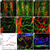
Figure 1
Figure 1. Time course of airway vessel changes after infection.
(A) Low-magnification view of highly ordered blood vessels (green) and lymphatics (red) in a flat whole mount of pathogen-free C57BL/6 mouse trachea. Most blood vessels and lymphatics are arranged in arcades in mucosa between cartilage rings. Capillaries of relatively uniform caliber (arrows) cross cartilage, but lymphatics do not. (B) Widened capillaries (arrows) over cartilages 7 days after M. pulmonis infection. (C) At 14 days, blood vessels are larger and lymphatic sprouts (arrows) are more abundant. Boxed regions enlarged in DF. (D) In pathogen-free mouse, LYVE-1 lymphatic sprouts are absent but some leukocytes (arrows) express LYVE-1. See also Supplemental Figure 1. (E) Vessel changes are accompanied by an influx of leukocytes, many stained for PECAM-1 (short arrows). Lymphatic sprouts are indicated by arrows. (F) Abundant lymphatic sprouts (arrows) and enlarged blood vessels. (G) Time course of changes in blood vessel diameter. *P < 0.05 versus pathogen-free. Data are represented as means ± SEM. (H) Triple-stained for PECAM-1, phosphohistone H3 (PH3), a marker of dividing nuclei, and LYVE-1 at 7 days. PH3-labeled endothelial nuclei (arrows) and nonendothelial cells (short arrows). (I) Cross-section of 14-day infected trachea. Many dividing nuclei (red) are present in epithelium and in other mucosal cells (short arrows). Although several labeled nuclei appear to coincide with lymphatics or blood vessels, most are actually superimposed and few (arrows) are truly located in vessels (arrows) as determined by examination of individual confocal sections. Scale bars: 200 μm (AC, H, and I); 50 μm (DF).
Figure 2
Figure 2. Expression of TNF family ligands and receptors in pathogen-free and M. pulmonis–infected mouse airways.
(A) qRT-PCR. Fold increase in mRNA compared with pathogen free. *P < 0.05 versus pathogen-free. Data are represented as means ± SEM. (BD) Confocal images of TNF-α and Iba1 immunoreactivity in cross-sections of (B and D) pathogen-free and (C and E) infected tracheas. (B) Strongest TNF-α staining is mainly in epithelium (arrows) in pathogen-free mouse. (C) Strong TNF-α staining is more widely distributed after infection. Some leukocytes (short arrows) are strongly stained. (D and E) Iba1, a marker of macrophages and dendritic cells, is sparse in pathogen-free trachea (D) but widely scattered in leukocytes after infection (E). Scale bar: 50 μm.
Figure 3
Figure 3. Distribution of TNF-R1 and lymphotoxin receptors on endothelial and periendothelial cells in tracheal whole mounts of pathogen-free mice.
(AC) TNF-R1 and (DF) LTBR immunoreactivity. Endothelial cells stained with rat anti-mouse PECAM-1 antibody and pericytes/smooth muscle cells stained with desmin and NG2 antibodies. Vessels: arterioles (A); capillaries (C); venules (V); lymphatics (L, faintly stained). TNF-R1 and LTBR immunoreactivity is stronger in venules and capillaries than in arterioles and very weak in lymphatics. Both TNF-R1 and LTBR staining have smooth continuous contours of endothelial cells rather than the sharp discrete outlines of periendothelial cells. Pericytes and vascular smooth muscle cells have very weak TNF-R1 and LBTR staining in contrast with strong staining of respiratory smooth muscle (see also Supplemental Figure 3). Scattered diffuse staining for TNR-R1 and LBTR in other mucosal cells, with strong LBTR staining in tracheal cartilages (*). For additional analysis of TNF-R1 staining combined with other markers of blood and lymphatic endothelial cells and periendothelial cells, see Supplemental Figure 3. Scale bar: 50 μm.
Figure 4
Figure 4. TNF-R1 immunoreactivity on endothelial cells of blood vessels.
(AF) Confocal images of pathogen-free mouse tracheas stained for TNF-R1 (red) and VE-cadherin (green) or (GI) for MECA-32 and LYVE-1. Strong TNF-R1 immunoreactivity of venules, intermediate staining of capillaries, and weak staining of arterioles and lymphatics. Boxed regions in AC are enlarged in DF. Inset in D shows punctate staining (arrows) of TNF-R1 in venular endothelial cells. (GI) TNF-R1 immunoreactivity is weak or absent in LYVE-1 immunoreactive (blue) lymphatics and nonendothelial cells (arrows). Scale bars: 200 μm (AC); 50 μm (DI); 25 μm (D, inset).
Figure 5
Figure 5. Blood vessel remodeling, lymphangiogenesis, and leukocyte influx after blocking TNF signaling.
(A) Extensive blood vessel remodeling and lymphangiogenesis in trachea of WT 14-day M. pulmonis–infected mouse. (B) Little vascular remodeling or lymphatic growth in infected Tnfr1–/– mouse. Scale bar: 200 μm. (C and D) Area density of blood vessels (C) and lymphatics (D) in WT mice treated with anti–TNF-α function–blocking antibody and in Tnfr1–/– (R1–/–) mice infected for 14 days. (E) Bronchial lymph node weight in anti–TNF-α antibody–treated WT mice and in infected Tnfr1–/– mice. P < 0.05, significantly different from (*) pathogen-free or (†) WT infected groups. Anti-TNF Ab, blocking antibody against mouse TNF-α. (F) qRT-PCR measurement of VEGF-A, -C, -D in tracheas of pathogen-free and 14-day infected WT and Tnfr1–/– mice. Expression of VEGF-C, but not VEGF-D is increased in infected WT mice, but not in infected Tnfr1–/– mice. VEGF-A expression is not increased in any infected airways. (GJ) H&E-stained sections of mouse lungs. (G) WT pathogen-free mouse has few H&E-stained leukocytes in lung parenchyma or airway lumen (*). (HJ) 14-day M. pulmonis–infected mice. (H) WT untreated mouse has extensive peribronchial cuffing and leukocytes in airway lumen (*) and lung parenchyma. Leukocyte influx is less prominent in infected WT mouse treated with TNF-α function–blocking antibody (I) and in infected Tnfr1–/– mouse (J). Scale bar: 50 μm.
Figure 6
Figure 6. Exaggerated blood vessel remodeling and lymphangiogenesis in infected Tnfα–/– mice.
(AC) Whole mount trachea of 14-day M. pulmonis–infected mouse stained for PECAM-1 (A) and LYVE-1 (B) showing numerous enlarged blood vessels (arrows) and newly formed lymphatics (short arrows) overlying cartilage rings. Numerous leukocytes expressing PECAM-1 are visible as small green dots. Images merged in C. Scale bar: 200 μm. (D and E) qRT-PCR analysis in pathogen-free and 3-day infected WT and corresponding Tnfα–/– mice. Copy number expressed relative to β-actin. (D) Ligands: TNF-α expression is not detected in Tnfα–/– mice. Expression of lymphotoxin A and B is increased in infected Tnfα–/– mice. (E) Receptors: TNF-R1 expression is increased in infected WT mice, and TNF-R2 is increased in infected WT and Tnfα–/– mice. *P < 0.05, significantly different from corresponding pathogen-free controls (n = 3 or more mice per group).
Figure 7
Figure 7. Lack of reversal of blood vessel remodeling and lymphangiogenesis after TNF-α blockade.
Measurements of blood vessels (A) and lymphatics (B) show that area densities increase during the first week after infection. Values for blood vessels reach a plateau, but those for lymphatics increase further during the second week. Anti–TNF-α antibody, given for 1 week beginning 1 week (1+1) after infection tends to reduce further growth of blood vessels or lymphangiogenesis but does not reverse it. Values for lymphatics tend to be less after addition of the anti–TNF-α antibody than after IgG, but the difference is not significant. *P < 0.05, significantly different from pathogen-free controls. (C) When anti–TNF-α antibody is given for 2 weeks beginning 4 weeks after infection (4+2), blood vessel remodeling and lymphangiogenesis is not significantly different from corresponding values after IgG injected during same period. (D) Weight of bronchial lymph nodes is less after anti–TNF-α antibody than after IgG and is almost reversed to values at the start of anti–TNF-α treatment. *P < 0.05, significantly different from start of anti–TNF-α treatment (n = 9–11 mice per group).

References

    1. Thurston G., Murphy T.J., Baluk P., Lindsey J.R., McDonald D.M. Angiogenesis in mice with chronic airway inflammation: strain-dependent differences. Am. J. Pathol. 1998;153:1099–1112. - PMC - PubMed
    1. Thurston G., Baluk P., McDonald D.M. Determinants of endothelial cell phenotype in venules. Microcirculation. 2000;7:67–80. doi: 10.1038/sj.mn.7300155. - DOI - PubMed
    1. Aurora A.B., et al. Immune complex-dependent remodeling of the airway vasculature in response to a chronic bacterial infection. J. Immunol. 2005;175:6319–6326. - PubMed
    1. Baluk P., et al. Functionally specialized junctions between endothelial cells of lymphatic vessels. J. Exp. Med. 2007;204:2349–2362. doi: 10.1084/jem.20062596. - DOI - PMC - PubMed
    1. Baluk P., et al. Pathogenesis of persistent lymphatic vessel hyperplasia in chronic airway inflammation. J. Clin. Invest. 2005;115:247–257. - PMC - PubMed

Publication types

MeSH terms

Substances