. 2009 Sep 17;4(9):e7089.
doi: 10.1371/journal.pone.0007089.

High resolution genome-wide analysis of chromosomal alterations in Burkitt's lymphoma

Affiliations

High resolution genome-wide analysis of chromosomal alterations in Burkitt's lymphoma

Saloua Toujani et al. PLoS One. .

Abstract

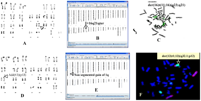
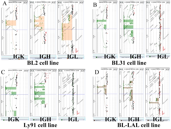
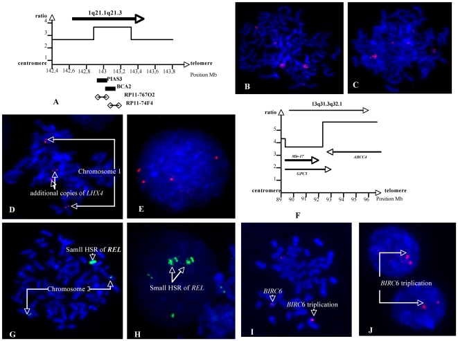
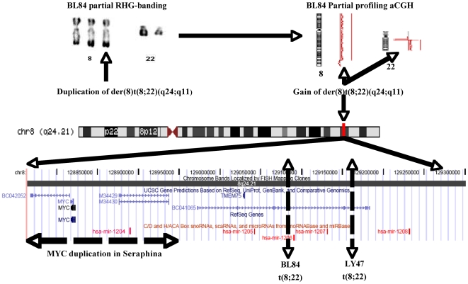
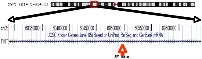
Additional chromosomal abnormalities are currently detected in Burkitt's lymphoma. They play major roles in the progression of BL and in prognosis. The genes involved remain elusive. A whole-genome oligonucleotide array CGH analysis correlated with karyotype and FISH was performed in a set of 27 Burkitt's lymphoma-derived cell lines and primary tumors. More than half of the 145 CNAs<2 Mb were mapped to Mendelian CNVs, including GSTT1, glutathione s-transferase and BIRC6, an anti-apoptotic protein, possibly predisposing to some cancers. Somatic cell line-specific CNVs localized to the IG locus were consistently observed with the 244 K aCGH platform. Among 136 CNAs >2 Mb, gains were found in 1q (12/27), 13q (7/27), 7q (6/27), 8q(4/27), 2p (3/27), 11q (2/27) and 15q (2/27). Losses were found in 3p (5/27), 4p (4/27), 4q (4/27), 9p (4/27), 13q (4/27), 6p (3/27), 17p (3/27), 6q (2/27),11pterp13 (2/27) and 14q12q21.3 (2/27). Twenty one minimal critical regions (MCR), (range 0.04-71.36 Mb), were delineated in tumors and cell lines. Three MCRs were localized to 1q. The proximal one was mapped to 1q21.1q25.2 with a 6.3 Mb amplicon (1q21.1q21.3) harboring BCA2 and PIAS3. In the other 2 MCRs, 1q32.1 and 1q44, MDM4 and AKT3 appeared as possible drivers of these gains respectively. The 13q31.3q32.1 <89.58-96.81> MCR contained an amplicon and ABCC4 might be the driver of this amplicon. The 40 Kb 2p16.1 <60.96-61> MCR was the smallest gained MCR and specifically encompassed the REL oncogene which is already implicated in B cell lymphomas. The most frequently deleted MCR was 3p14.1 <60.43-60.53> that removed the fifth exon of FHIT. Further investigations which combined gene expression and functional studies are essential to understand the lymphomagenesis mechanism and for the development of more effective, targeted therapeutic strategies.


Conflict of interest statement

Competing Interests: The authors have declared that no competing interests exist.

Figures

Figure 1
Figure 1. Correlation karyotype-high resolution aCGH.
A RHG-banded karyotype of Ly47 showing 2 normal chromosome 16 (arrow). B Karyogramme of Ly47 cell line showing a gain of chromosome 16 (arrow). C FISH with whole painting of chromosome 16 confirmed aCGH result. The additional piece of chromosome 16 was translocated at the long arm of chromosome 11. D RHG-banded karyotype of Ly91 showing an add(13)(p12) which is presented in 20% of cells (arrow). E Karyogramme of Ly91 cell line showing a non segmented gain on 1q (arrow). F FISH with whole painting of chromosome 1(red) and 13(green) allowed to identify the origin of the material translocated at 13p. Hence, the add(13) became a der (13)t(1;13)(q32.1;p12).
Figure 2
Figure 2. Immunoglobulin Somatic Copy Number Variation.
The immunoglobulin loci at chromosomal subbands 2p11.2 (IGK), 14q32.3 (IGH) and 22q11.2 (IGL) showed acquired monoclonal alterations by the 244 K aCGH platform. The segmented rearrangements were identified by a stained rectangle and the individual oligonucleotides are identified by a bold dot. The Y axis is the chromosomal position, the X axis is the ratio (loss is to the left). A to D, S1: The IGH locus was rearranged in all cell lines (14/15) on the two chromosomes, with a specific pattern for each cell line. BL2 and BL31 have a t(8;14). A to D, S1: IGK gene rearrangements were detected in all cell lines and 8 have a clear biallelic rearrangement including LY91 the only t(2;8). A to D, S1: IGL loci somatic rearrangements were observed in only 10 cell lines (7 biallelics) including four cases of t(8;22) as in BLAL. Other cell lines are presented as supporting figures.
Figure 3
Figure 3. Various gained or amplified small regions description.
A. Copy number profile of 1q21.1 amplicon in BL41:the 7 copies maximal amplitude region was the <142,87–143,28 Mb> interval that included BCA2 and PIAS3 genes. B and C. The amplifications of BCA2 and PIAS3 were confirmed by FISH with BAC clones RP11-767O2 and RP11-74F4 respectively which showed a small HSR. D and E. In Namalwa a low level amplification (4 to 5 copies) of 1q25.2 <176,6–177> was confirmed by FISH with BAC clone RP11-175C8 which contains the LHX4 gene only. 3F. In Namalwa the 13q31.33q2.1 <89,58–95,82> region was amplified. The proximal part that harboured the polycistron mir-17 and a part of GPC5 gene was present in 8 copies. The distal region, with maximal amplitude (12 copies), contained 12 genes including GPC5, GPC6 and ABCC4 (see text). G and H. In BL 41 the REL gene was found amplified and FISH with BAC clone RP11-373L24 showed a small HSR. I and J Molecular cytogenetic on BLMer cell line showing a duplication of BIRC6. RP11-121M15 BAC clone was choosen on the region of BIRC6. The latter gene was duplicated as denoted by an arrow.
Figure 4
Figure 4. Virtual cloning of 8q24 region.
MYC region breakpoints. In Seraphina, BL 84 and Ly 47 cell lines, MYC locus was gained. MYC region breakpoints vary with primary anomalies. In BL 84 and Ly 47 cell lines with t(8;22), MYC breakpoints were mapped to PVT1(Huppi et al., 2008); mir-1205 and mir1204 (not present on UCSC map, located on chr8:128877390-128877456) defined the MCR of this gain. In Seraphina, a 240 Kb duplication was observed around MYCand located by FISH in the immediate vicinity this gene on the der(14)t(8;14) (data not shown). This duplication contained hsa mir 1204.
Figure 5
Figure 5. A small intragenic loss.
Based on the UCSC data base (June, 2004), the 402 Kb MCR on 3p remove the fifth exon of FHIT gene.

References

    1. Harris NL, Horning SJ. Burkitt's lymphoma–the message from microarrays. N Engl J Med. 2006;354:2495–2498. - PubMed
    1. Thorley-Lawson D, Allday M. The curious case of the tumour virus: 50 years of Burkitt's lymphoma. Nat Rev Microbiol. 2008;6:913–924. - PubMed
    1. Lenoir G, Preud'homme J, Bernheim A, Berger R. Correlation between immunoglobulin light chain expression and variant translocation in Burkitt's lymphoma. Nature. 1982;298:474–476. - PubMed
    1. Hummel M, Bentink S, Berger H, Klapper W, Wessendorf S, et al. A biologic definition of Burkitt's lymphoma from transcriptional and genomic profiling. N Engl J Med. 2006;354:2419–2430. - PubMed
    1. Battey J, Moulding C, Taub R, Murphy W, Stewart T, et al. The human c-myc oncogene: structural consequences of translocation into the IgH locus in Burkitt lymphoma. Cell. 1983;34:779–787. - PubMed

Publication types