. 2009 Sep 16;4(9):e6964.
doi: 10.1371/journal.pone.0006964.

Something old, something new, something borrowed; how the thermoacidophilic archaeon Sulfolobus solfataricus responds to oxidative stress

Affiliations

Something old, something new, something borrowed; how the thermoacidophilic archaeon Sulfolobus solfataricus responds to oxidative stress

Walid S Maaty et al. PLoS One. .

Abstract

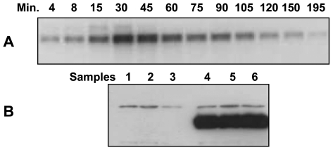
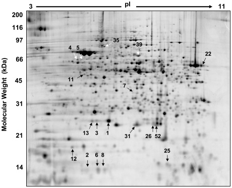
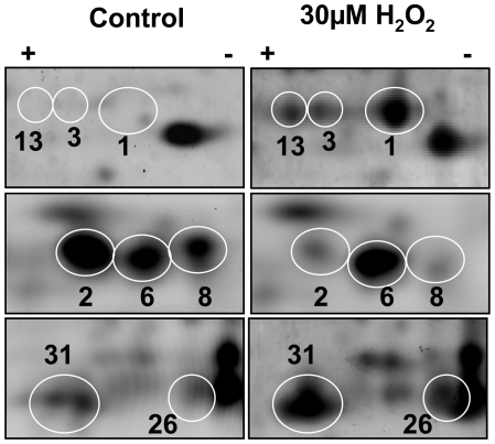
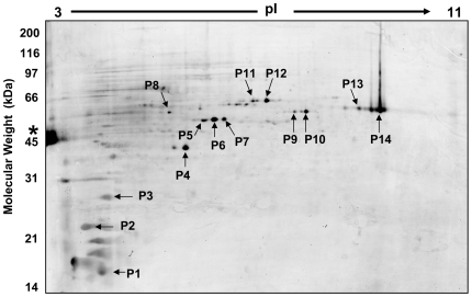
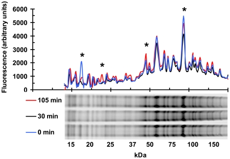
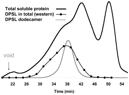
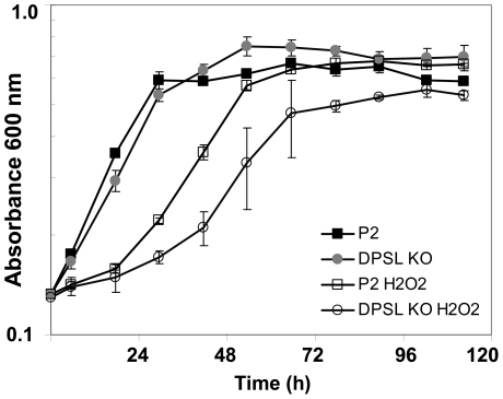
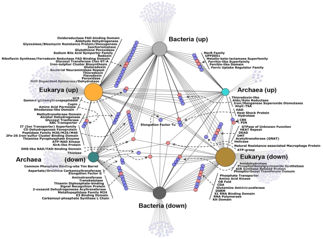
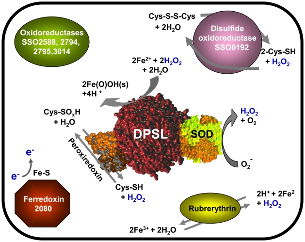
To avoid molecular damage of biomolecules due to oxidation, all cells have evolved constitutive and responsive systems to mitigate and repair chemical modifications. Archaea have adapted to some of the most extreme environments known to support life, including highly oxidizing conditions. However, in comparison to bacteria and eukaryotes, relatively little is known about the biology and biochemistry of archaea in response to changing conditions and repair of oxidative damage. In this study transcriptome, proteome, and chemical reactivity analyses of hydrogen peroxide (H(2)O(2)) induced oxidative stress in Sulfolobus solfataricus (P2) were conducted. Microarray analysis of mRNA expression showed that 102 transcripts were regulated by at least 1.5 fold, 30 minutes after exposure to 30 microM H(2)O(2). Parallel proteomic analyses using two-dimensional differential gel electrophoresis (2D-DIGE), monitored more than 800 proteins 30 and 105 minutes after exposure and found that 18 had significant changes in abundance. A recently characterized ferritin-like antioxidant protein, DPSL, was the most highly regulated species of mRNA and protein, in addition to being post-translationally modified. As expected, a number of antioxidant related mRNAs and proteins were differentially regulated. Three of these, DPSL, superoxide dismutase, and peroxiredoxin were shown to interact and likely form a novel supramolecular complex for mitigating oxidative damage. A scheme for the ability of this complex to perform multi-step reactions is presented. Despite the central role played by DPSL, cells maintained a lower level of protection after disruption of the dpsl gene, indicating a level of redundancy in the oxidative stress pathways of S. solfataricus. This work provides the first "omics" scale assessment of the oxidative stress response for an archeal organism and together with a network analysis using data from previous studies on bacteria and eukaryotes reveals evolutionarily conserved pathways where complex and overlapping defense mechanisms protect against oxygen toxicity.


Conflict of interest statement

Competing Interests: The authors have declared that no competing interests exist.

Figures

Figure 1
Figure 1. Treatment of S. solfataricus with 30 µM H2O2 leads to an up-regulation in transcription and translation of DPSL.
A) Time course northern blot analysis for dpsl mRNA after treatment with 30 µM H2O2. B) Western blots for DPSL, 105 minutes after addition of 30 µM H2O2. Lanes 1–3 controls, 4–6 are from the three biological replicates used for the microarray and proteomics experiments. The polyclonal antibody to DPSL protein recognizes a background protein of slightly greater molecular weight than DPSL.
Figure 2
Figure 2. Regulated proteins of the S. solfataricus proteome after exposure to 30 µM H2O2.
Approximately 818 spots (common to all gels) were used in the CyDye 2D DIGE analysis. 18 protein spots changed significantly in abundance 105 minutes post H2O2. Spots that changed in abundance are indicated with arrows. Protein identifications were made using in-gel proteolysis followed by LC-MS/MS and are listed in Table 2.
Figure 3
Figure 3. Post translational modification is a common feature in S. solfataricus.
Three of the proteins that are regulated after H2O2 treatment are known to be important in oxidative stress and were found in multiple gel spots. Modifications can alter the pI and MW position on 2D gels. The paired panels show close ups of SyproRuby stained 2D gels, 105 minutes after addition of 30 µM H2O2. Top, DPSL (SSO2079; 21639 Da; pI 5.25) spots 1, 3 and 13. Middle, Rubrerythrin (SSO2642; 16081 Da; pI 5.44) spots 2, 6 and 8. Bottom, Peroxiredoxin (SSO2121; 24786Da; pI 6.85) spots 26 and 31.
Figure 4
Figure 4. 2D gel of the S. solfataricus phospho-proteome 105 minutes after H2O2 treatment.
The gel was stained with phosphoprotein specific stain ProQ Diamond. Each of the numbered spots was picked and the proteins were identified using in-gel proteolysis followed LC-MS/MS. Ovalbumin (* on left) is a 45 kDa phosphoprotein standard.
Figure 5
Figure 5. Protein thiol reactivity changes in H2O2 stressed S. solfataricus.
Proteome-wide labeling of free cysteine thiols, with BODIPY maleimide, shows that there is a population of redox sensitive proteins. Plot on the top shows the average fluorescent signal with respect to molecular weight. The fluorescent signal from three experiments was combined and normalized for total protein. The gel lanes at the bottom show the actual data form one experiment; with 0 (blue line), 30 (black line) and 105 minute (red line) samples. * indicates protein bands that were highly sensitive to changes in redox potential.
Figure 6
Figure 6. Size exclusion chromatography of DPSL.
SEC data shows that a significant portion of DPSL from H2O2 stressed cells is part of a larger molecular complex. Total soluble protein, 105 min. after H2O2 exposure (black line) and purified recombinant DPSL separated under identical conditions (gray line) were detected by monitoring at 280 nm. Western blot analysis of the total soluble protein fractions using anti-DPSL antibody shows that in vivo part of the DPSL elutes earlier (27–34 min) in comparison to the purified wild type DPSL (∼38 min) indicating that it is part of a larger molecular complex.
Figure 7
Figure 7. DPSL deficient strain of S. solfataricus is more sensitive to H2O2.
S. solfataricus (P2) and a mutant lacking DPSL (DPSL KO) were cultured with and without 30 µM H2O2. P2 (solid square), P2 with H2O2 (open square), DPSL KO (gray circle), DPSL KO with H2O2 (open circle) n = 3.
Figure 8
Figure 8. Network of shared mechanisms for oxidative stress response between Archaea, Bacteria, and Eukaryotes.
Data from transcriptomics and proteomics experiments on S. solfataricus, Bacillus, E. coli, and Yeast after H2O2 exposure were combined to assess the relatedness of representative organisms across the three domains of life. Blue nodes represent protein families and salmon nodes represent protein clans. Smaller gray nodes show pfams unique to a particular domain and direction of regulation. The size of the node for each domain is scaled to according to number of regulated pfams.
Figure 9
Figure 9. Schematic of the oxidative stress response in S. solfataricus based on mRNA and protein regulation after hydrogen peroxide exposure.
Numbers indicate gene number (SSO). DPSL, SOD, and Peroxiredoxin are part of a molecular complex that can coordinate removal of ROS by converting highly reactive superoxide into H2O2 and then using this as substrate in subsequent reactions.

References

    1. Imlay JA. Pathways of oxidative damage. Annual Review of Microbiology. 2003;57:395–418. - PubMed
    1. Gutteridge JMC, Halliwell B. Free radicals and antioxidants in the year 2000 - A historical look to the future. Reactive Oxygen Species: From Radiation to Molecular Biology. 2000;899:136–147. - PubMed
    1. Slupphaug G, Kavli B, Krokan HE. The interacting pathways for prevention and repair of oxidative DNA damage. Mutation Research-Fundamental and Molecular Mechanisms of Mutagenesis. 2003;531:231–251. - PubMed
    1. Farr SB, Kogoma T. Oxidative Stress Responses in Escherichia-Coli and Salmonella-Typhimurium. Microbiological Reviews. 1991;55:561–585. - PMC - PubMed
    1. Power JHT, Asad S, Chataway TK, Chegini F, Manavis J, et al. Peroxiredoxin 6 in human brain: molecular forms, cellular distribution and association with Alzheimer's disease pathology. Acta Neuropathologica. 2008;115:611–622. - PMC - PubMed

Publication types

MeSH terms