. 2009 Sep 17;4(9):e7074.
doi: 10.1371/journal.pone.0007074.

PSSA-2, a membrane-spanning phosphoprotein of Trypanosoma brucei, is required for efficient maturation of infection

Affiliations

PSSA-2, a membrane-spanning phosphoprotein of Trypanosoma brucei, is required for efficient maturation of infection

Cristina M Fragoso et al. PLoS One. .

Abstract

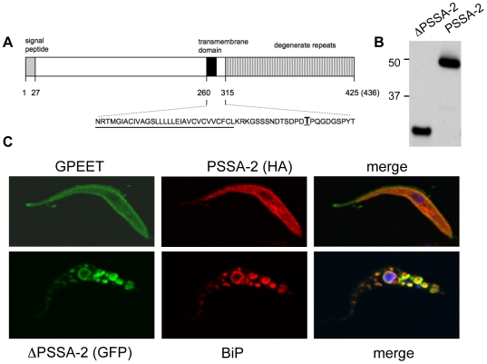
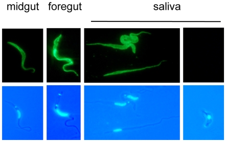
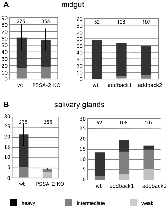
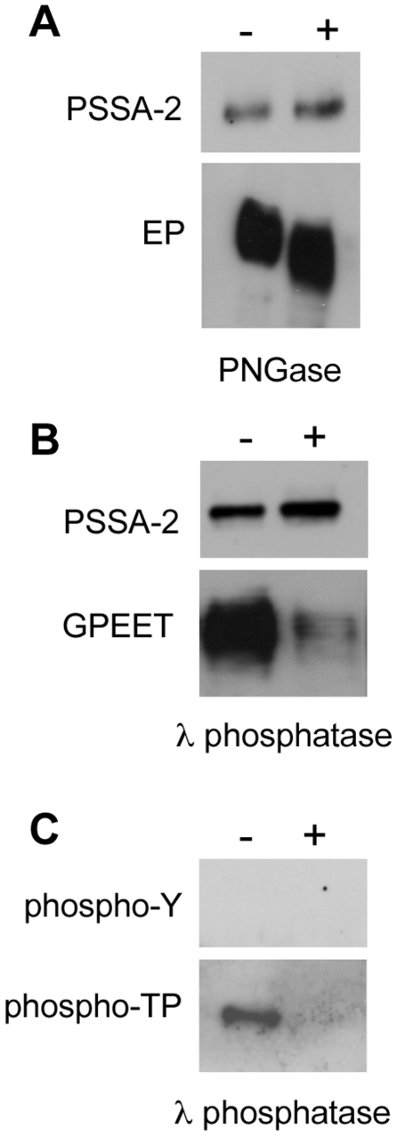
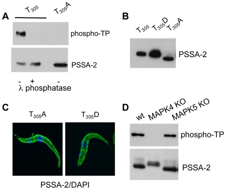
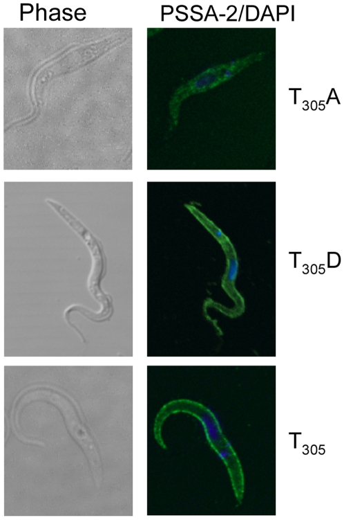
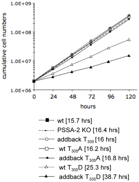
The coat of Trypanosoma brucei consists mainly of glycosylphosphatidylinositol-anchored proteins that are present in several million copies and are characteristic of defined stages of the life cycle. While these major components of the coats of bloodstream forms and procyclic (insect midgut) forms are well characterised, very little is known about less abundant stage-regulated surface proteins and their roles in infection and transmission. By creating epitope-tagged versions of procyclic-specific surface antigen 2 (PSSA-2) we demonstrated that it is a membrane-spanning protein that is expressed by several different life cycle stages in tsetse flies, but not by parasites in the mammalian bloodstream. In common with other membrane-spanning proteins in T. brucei, PSSA-2 requires its cytoplasmic domain in order to exit the endoplasmic reticulum. Correct localisation of PSSA-2 requires phosphorylation of a cytoplasmic threonine residue (T(305)), a modification that depends on the presence of TbMAPK4. Mutation of T(305) to alanine (T(305)A) has no effect on the localisation of the protein in cells that express wild type PSSA-2. In contrast, this protein is largely intracellular when expressed in a null mutant background. A variant with a T(305)D mutation gives strong surface expression in both the wild type and null mutant, but slows growth of the cells, suggesting that it may function as a dominant negative mutant. The PSSA-2 null mutant exhibits no perceptible phenotype in culture and is fully competent at establishing midgut infections in tsetse, but is defective in colonising the salivary glands and the production of infectious metacyclic forms. Given the protein's structure and the effects of mutation of T(305) on proliferation and localisation, we postulate that PSSA-2 might sense and transmit signals that contribute to the parasite's decision to divide, differentiate or migrate.


Conflict of interest statement

Competing Interests: The authors have declared that no competing interests exist.

Figures

Figure 1
Figure 1. PSSA-2 requires the cytoplasmic tail for surface localisation.
A. Schematic representation of PSSA-2. Predicted structural domains and signal peptide are indicated (not drawn to scale). AnTat 1.1 encodes a polypeptide of 436 amino acids, whereas the genome strain TREU 927/4 encodes a polypeptide of 425 amino acids. The sequence corresponding to the transmembrane domain is underlined; threonine residue T305 is in boldface type and underlined. B. Immunoblot analysis of total lysates from procyclic forms of AnTat 1.1 stably transfected with plasmids encoding either an HA-tagged version of truncated PSSA-2 (ΔPSSA-2), lacking the cytoplasmic domain from residues 292–436, or full-length PSSA-2. Proteins were detected with an anti-HA antibody. 106 cell equivalents were loaded per lane. Markers are indicated on the left. C. Immunofluorescence analysis of HA-tagged full length PSSA-2 (top panel) and a ΔPSSA-2/GFP fusion protein (lower panel). Trypanosomes were fixed with formaldehyde and glutaraldehyde, permeabilized with Triton X-100 and stained with anti-GPEET, anti-HA or anti-BiP antibodies as indicated.
Figure 2
Figure 2. Detection of PSSA-2 and procyclin during synchronous differentiation of stumpy bloodstream forms to procyclic forms.
A. Northern blot analysis of wild type AnTat 1.1. Numbers denote hours after triggering differentiation. +G: early procyclic forms cultured in the presence of glycerol. −G: late procyclic forms cultured without glycerol. EtBr: ethidium bromide-stained rRNA is shown as a loading control. B. Immunofluorescence analysis of cells expressing HA-tagged PSSA-2 during differentiation from bloodstream forms to procyclic forms. At given time points after differentiation was triggered, cells were fixed and stained with DAPI (left panels), anti-HA to detect PSSA-2 (central panels) and anti-EP (right panels).
Figure 3
Figure 3. Expression of PSSA-2 in tsetse-derived trypanosomes.
Immunofluorescence analysis of PSSA-2-HA expression by trypanosomes isolated from the midgut, foregut and saliva probes 28–35 days after the infective bloodmeal. Trypanosomes were fixed with formaldehyde and glutaraldehyde and stained with anti-HA antibody (upper panel) and DAPI (lower panel).
Figure 4
Figure 4. PSSA-2 does not influence midgut infections in tsetse, but is required for efficient colonisation of the salivary glands.
Teneral flies were infected with procyclic forms of AnTat 1.1 (wild type), the PSSA-2 null mutant (PSSA-2 KO) or the independent addback clones 1 and 2. Flies were dissected 28–35 days post infection and graded for the prevalence and intensity of infections in the midgut and salivary glands. The y-axis represents the percentage of infected flies. The total number of flies is indicated above the bars on the graphs. For the panel on the left, this represents the total numbers from 4 independent experiments. Transmission index (see text) is the percentage of midgut infections giving rise to salivary gland infections.
Figure 5
Figure 5. Post-translational modification of PSSA-2.
A. Total lysates from procyclic forms stably expressing HA-tagged PSSA-2 were incubated in the presence (+) or absence (−) of PNGase and analysed by immunoblotting. Top panel: PSSA-2 detected with anti-HA ; lower panel, anti-EP. B. Total lysates from procyclic forms stably expressing HA-tagged PSSA-2 were incubated in the presence (+) or absence (−) of λ phosphatase and analysed by immunoblotting. MAb 5H3 (second panel) preferentially binds phosphorylated GPEET . C. HA-tagged PSSA-2 was immunoprecipitated, incubated with (+) or without (−) λ phosphatase and detected with antibodies against phosphotyrosine (phosphoY; upper panel) or phosphothreonine-proline (phospho-TP; lower panel).
Figure 6
Figure 6. Confirmation that T305 is phosphorylated.
A. HA-tagged PSSA-2 or PSSA-2(T305A) were immunoprecipated, incubated in the presence (+) or absence (−) of λ phosphatase and analysed by immunoblotting with antibodies against phospho-TP (upper panel) or anti-HA, to detect PSSA-2 (lower panel). B. Mutation of T305 to alanine alters the electrophoretic mobility of the protein. Total lysates from cells stably expressing PSSA-2(T305), PSSA-2(T305A) or PSSA-2(T305D) were detected with anti-HA antibodies. 1.5×10⁵ cell equivalents were loaded in the first two lanes and 3×10⁶ for PSSA-2(T305A), which is much more weakly expressed. C. PSSA-2(T305A) and PSSA-2(T305D) localise to the plasma membrane. Cells were fixed and permeablised as described in the legend to Figure 1. D. MAP kinase 4 is required for phosphorylation of T305. Null mutants of MAPK4 (MAPK4 KO) and MAPK5 (MAPK5 KO) were stably transformed with HA-tagged PSSA-2. The protein was immunoprecipated and detected with anti-phospho-TP antibodies to detect phospho-T305 or anti-HA to detect PSSA-2.
Figure 7
Figure 7. Surface localisation of PSSA-2(T305A) depends on the genetic background.
HA-tagged wild type PSSA-2(T305) or the mutant forms T305A or T305D were expressed in a PSSA-2 null mutant. Left panel, phase contrast; right panel, detection of PSSA-2 with anti-HA antibodies/DAPI staining of DNA. Cells were fixed and permeablised as described in the legend to Figure 1.
Figure 8
Figure 8. Mutation of T305 to aspartic acid has a deleterious effect on growth.
Comparison of the population doubling time of wild type AnTat 1.1 with various PSSA-2 mutants. Cell densities were adjusted daily to 2×10⁶ cells ml−1 in order to ensure logarithmic growth. Generation times are shown in square brackets.

References

    1. Vassella E, Den Abbeele JV, Bütikofer P, Renggli CK, Furger A, et al. A major surface glycoprotein of Trypanosoma brucei is expressed transiently during development and can be regulated post-transcriptionally by glycerol or hypoxia. Genes Dev. 2000;14:615–626. - PMC - PubMed
    1. Urwyler S, Studer E, Renggli CK, Roditi I. A family of stage-specific alanine-rich proteins on the surface of epimastigote forms of Trypanosoma brucei. Mol Microbiol. 2007;63:218–228. - PubMed
    1. Hajduk SL. Antigenic variation during the developmental cycle of Trypanosoma brucei. J Protozool. 1984;31:41–47. - PubMed
    1. Utz S, Roditi I, Kunz Renggli C, Almeida IC, Acosta-Serrano A, et al. Trypanosoma congolense Procyclins: Unmasking Cryptic Major Surface Glycoproteins in Procyclic Forms. Eukaryot Cell. 2006;5:1430–1440. - PMC - PubMed
    1. Bayne RA, Kilbride EA, Lainson FA, Tetley L, Barry JD. A major surface antigen of procyclic stage Trypanosoma congolense. Mol Biochem Parasitol. 1993;61:295–310. - PubMed

Publication types

MeSH terms

Substances

LinkOut - more resources