

Spontaneous and radiation-induced chromosomal instability and persistence of chromosome aberrations after radiotherapy in lymphocytes from prostate cancer patients
- PMID: 19760427
- PMCID: PMC2822223
- DOI: 10.1007/s00411-009-0244-x
Spontaneous and radiation-induced chromosomal instability and persistence of chromosome aberrations after radiotherapy in lymphocytes from prostate cancer patients
Abstract
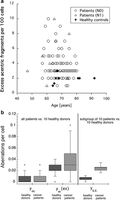
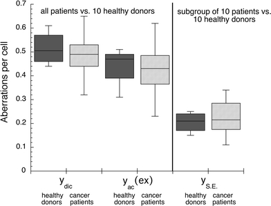
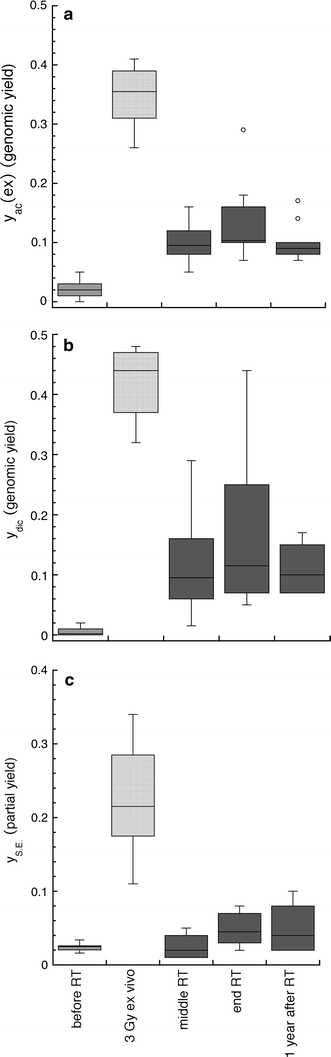
The aim of the study was to compare the spontaneous and ex vivo radiation-induced chromosomal damage in lymphocytes of untreated prostate cancer patients and age-matched healthy donors, and to evaluate the chromosomal damage, induced by radiotherapy, and its persistence. Blood samples from 102 prostate cancer patients were obtained before radiotherapy to investigate the excess acentric fragments and dicentric chromosomes. In addition, in a subgroup of ten patients, simple exchanges in chromosomes 2 and 4 were evaluated by fluorescent in situ hybridization (FISH), before the onset of therapy, in the middle and at the end of therapy, and 1 year later. Data were compared to blood samples from ten age-matched healthy donors. We found that spontaneous yields of acentric chromosome fragments and simple exchanges were significantly increased in lymphocytes of patients before onset of therapy, indicating chromosomal instability in these patients. Ex vivo radiation-induced aberrations were not significantly increased, indicating proficient repair of radiation-induced DNA double-strand breaks in lymphocytes of these patients. As expected, the yields of dicentric and acentric chromosomes, and the partial yields of simple exchanges, were increased after the onset of therapy. Surprisingly, yields after 1 year were comparable to those directly after radiotherapy, indicating persistence of chromosomal instability over this time. Our results indicate that prostate cancer patients are characterized by increased spontaneous chromosomal instability. This instability seems to result from defects other than a deficient repair of radiation-induced DNA double-strand breaks. Radiotherapy-induced chromosomal damage persists 1 year after treatment.
Figures
Similar articles
-
Chromosomal instability in peripheral blood lymphocytes and risk of prostate cancer.Cancer Epidemiol Biomarkers Prev. 2005 Mar;14(3):748-52. doi: 10.1158/1055-9965.EPI-04-0236. Cancer Epidemiol Biomarkers Prev. 2005. PMID: 15767363
-
Chromosomal in-vitro radiosensitivity of lymphocytes in radiotherapy patients and AT-homozygotes.Strahlenther Onkol. 1998 Oct;174(10):510-6. doi: 10.1007/BF03038983. Strahlenther Onkol. 1998. PMID: 9810318
-
Biodosimetry Based on γ-H2AX Quantification and Cytogenetics after Partial- and Total-Body Irradiation during Fractionated Radiotherapy.Radiat Res. 2015 Apr;183(4):432-46. doi: 10.1667/RR13911.1. Epub 2015 Apr 6. Radiat Res. 2015. PMID: 25844946
-
BRCA1 role in the mitigation of radiotoxicity and chromosomal instability through repair of clustered DNA lesions.Chem Biol Interact. 2010 Nov 5;188(2):350-8. doi: 10.1016/j.cbi.2010.03.046. Epub 2010 Apr 4. Chem Biol Interact. 2010. PMID: 20371364 Review.
-
Studies on chromosome aberration induction: what can they tell us about DNA repair?DNA Repair (Amst). 2006 Sep 8;5(9-10):1171-81. doi: 10.1016/j.dnarep.2006.05.033. Epub 2006 Jun 30. DNA Repair (Amst). 2006. PMID: 16814619 Review.
Cited by
-
Investigation of Chromosome 1 Aberrations in the Lymphocytes of Prostate Cancer and Benign Prostatic Hyperplasia Patients by Fluorescence in situ Hybridization.Cancer Manag Res. 2021 May 31;13:4291-4298. doi: 10.2147/CMAR.S293249. eCollection 2021. Cancer Manag Res. 2021. PMID: 34103984 Free PMC article.
-
Numerical chromosomal instability mediates susceptibility to radiation treatment.Nat Commun. 2015 Jan 21;6:5990. doi: 10.1038/ncomms6990. Nat Commun. 2015. PMID: 25606712 Free PMC article.
-
Dose-Effects Models for Space Radiobiology: An Overview on Dose-Effect Relationships.Front Public Health. 2021 Nov 8;9:733337. doi: 10.3389/fpubh.2021.733337. eCollection 2021. Front Public Health. 2021. PMID: 34820349 Free PMC article. Review.
-
Why it is crucial to analyze non clonal chromosome aberrations or NCCAs?Mol Cytogenet. 2016 Feb 13;9:15. doi: 10.1186/s13039-016-0223-2. eCollection 2016. Mol Cytogenet. 2016. PMID: 26877768 Free PMC article.
-
DNA repair deficiency in peripheral blood lymphocytes of endometrial cancer patients with a family history of cancer.BMC Cancer. 2014 Oct 15;14:765. doi: 10.1186/1471-2407-14-765. BMC Cancer. 2014. PMID: 25315979 Free PMC article.
References
-
- Arsova-Sarafinovska Z, Eken A, Matevska N, Erdem O, Sayal A, Savaser A, Banev S, Petrovski D, Dzikova S, Georgiev V, Sikole A, Ozgok Y, Suturkova L, Dimovski AJ, Aydin A (2009) Increased oxidative/nitrosative stress and decreased antioxidant enzyme activities in prostate cancer. Clin Biochem (Epub ahead of print) - PubMed
Publication types
MeSH terms
LinkOut - more resources
Full Text Sources
Medical
Research Materials
