Comparative Study
. 2009 Oct 13;48(40):9556-68.
doi: 10.1021/bi901110n.

Biophysical characterization of the iron in mitochondria from Atm1p-depleted Saccharomyces cerevisiae

Affiliations
Comparative Study

Biophysical characterization of the iron in mitochondria from Atm1p-depleted Saccharomyces cerevisiae

Ren Miao et al. Biochemistry. .

Abstract

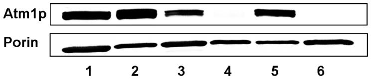
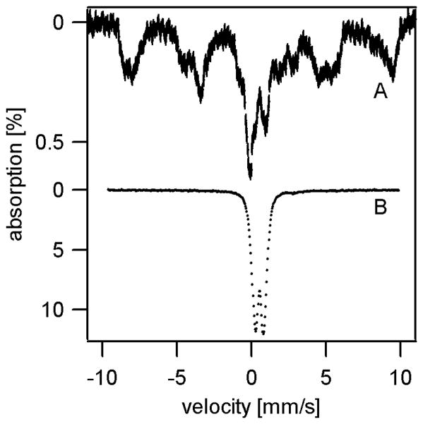
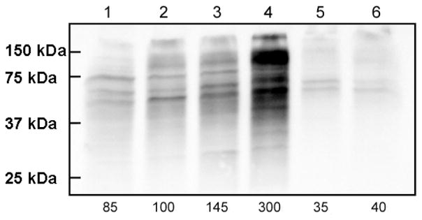
Atm1p is an ABC transporter localized in the mitochondrial inner membrane; it functions to export an unknown species into the cytosol and is involved in cellular iron metabolism. Depletion or deletion of Atm1p causes Fe accumulation in mitochondria and a defect in cytosolic Fe/S cluster assembly but reportedly not a defect in mitochondrial Fe/S cluster assembly. In this study the nature of the accumulated Fe was examined using Mossbauer spectroscopy, EPR, electronic absorption spectroscopy, X-ray absorption spectroscopy, and electron microscopy. The Fe that accumulated in aerobically grown cells was in the form of iron(III) phosphate nanoparticles similar to that which accumulates in yeast frataxin Yfh1p-deleted or yeast ferredoxin Yah1p-depleted cells. Relative to WT mitochondria, Fe/S cluster and heme levels in Atm1p-depleted mitochondria from aerobic cells were significantly diminished. Atm1p depletion also caused a buildup of nonheme Fe(II) ions in the mitochondria and an increase in oxidative damage. Atm1p-depleted mitochondria isolated from anaerobically grown cells exhibited WT levels of Fe/S clusters and hemes, and they did not hyperaccumulate Fe. Atm1p-depleted cells lacked Leu1p activity, regardless of whether they were grown aerobically or anaerobically. These results indicate that Atm1p does not participate in mitochondrial Fe/S cluster assembly and that the species exported by Atm1p is required for cytosolic Fe/S cluster assembly. The Fe/S cluster defect and the Fe-accumulation phenotype, resulting from the depletion of Atm1p in aerobic cells (but not in anaerobic cells), may be secondary effects that are observed only when cells are exposed to oxygen during growth. Reactive oxygen species generated under these conditions might degrade iron-sulfur clusters and lower heme levels in the organelle.


Figures

Figure 1
Figure 1
Western blots of Atm1p and porin in isolated mitochondria. Lane 1, W303/Gal/O2; Lane 2, W303/Glu/O2; Lane 3, Gal-ATM1/Gal/O2; Lane 4, Gal-ATM1/Glu/O2; Lane 5, W303/Glu/Ar; Lane 6, Gal-ATM1/Glu/Ar. Top panel, blot treated with anti-Atm1p antibody; Bottom panel, blot treated with an antibody for the mitochondrial outer membrane protein porin which served as a loading control.
Figure 2
Figure 2
4.5 K Mössbauer spectra of mitochondria from Gal-ATM1 and W303 cells. A, Gal-ATM1/Glu/O2, collected with 40 mT field applied parallel to gamma rays (low applied field). The solid red line is the sum of the Fe(III) nanoparticle doublet (δ = 0.53 mm/s, ΔEQ = 0.73 mm/s) and a HS Fe(II) doublet; B, same as A, but with 8 T applied field; C, Gal-ATM1/Gal/O2, low applied field. The solid red line is the sum of the Fe(III) nanoparticle double (δ = 0.52 mm/s, Δ EQ = 0.73 mm/s) and a HS Fe(II) doublet; D, same as A but reduced with sodium dithionite; E, same as C but reduced with sodium dithionite. The solid line above the data outlines a [Fe4S4]2+ doublet (δ = 0.45 mm/s, ΔEQ = 1.15 mm/s); F, W303/Glu/Ar, low applied field, the solid line above the data outlines the sum of two major doublets ([Fe4S4]2+ and HS Fe(II)), representing ~ 70% of Fe; G, Gal-ATM1/Glu/Ar, low applied field. The solid line above the data outlines the sum of two major doublets, representing ~ 80% of Fe.
Figure 3
Figure 3
X-band EPR spectra of isolated mitochondria from Gal-ATM1 and W303 cells. Top panel, from Gal-ATM1/Glu/O2 cells. A, 10 K; B, 30 K; C, 80 K; microwave frequency for all spectra, 9.46 GHz; microwave power for A, B and C, 20 mW. Middle panel, g = 2 region. A, W303/Gal/O2; B, Gal-ATM1/Glu/O2; C, same as B, but reduced with sodium dithionite; D, W303/Glu/Ar; E, Gal-ATM1/Glu/Ar; temperature. Temperature, 10 K, microwave power, 0.2 mW. Bottom panel, g = 4–6 region. A, as-isolated Gal-ATM1/Glu/O2 mitochondria; B, same as A but treated with sodium dithionite. Temperature, 10 K; microwave power, 20 mW.
Figure 4
Figure 4
STEM-HAADF images and elemental maps of iron deposits in mitochondria isolated from Gal-ATM1/Glu/O2 cells. Images I, II, and II were obtained at increasing magnification, with the red box in each panel corresponding approximately to the full image in the next lower panel. The red box in image I also corresponds to the region scanned for Fe, P, O, and C maps. The bright spots around the center and the corner of the carbon map are probably a beam-damaged mark.
Figure 5
Figure 5
X-ray absorption spectra of mitochondria isolated from Gal-ATM1/Glu/O2 cells. a, XANES; b, FT (k = 2.0 – 16.0 Å−1, k3 weighting); c, EXAFS. Solid red lines are simulations assuming the FeO5P2 model of Table 1.
Figure 6
Figure 6
Electronic absorption reduced-minus-oxidized difference spectra of mitochondria isolated from W303 and Gal-ATM1 cells. Spectra were obtained at room temperature from samples treated with sodium dithionite. They were subtracted from those obtained from samples treated with ferricyanide. A, W303/Glu/O2; B, Gal-ATM1/Glu/O2; C, from W303/Glu/Ar; D, Gal-ATM1/Glu/Ar.
Figure 7
Figure 7
Mössbauer spectra showing high-spin Fe(II) species in Atm1p-depleted and Yah1p-depleted mitochondria and whole cells. All spectra were collected 4.5 K with 40 mT parallel applied field. A, Gal-ATM1/Glu/O2 mitochondria; B, Gal-ATM1/Gal/O2 mitochondria; C, Gal-ATM1/Glu/O2 cells; D, mitochondria from Yah1p-depleted cells grown on glucose/O2 (batch 1); E, Same as B, but a different batch harvested after 35 hr of growth. A vertical dashed line is drawn at 2.75 mm/s.
Figure 8
Figure 8
4.5 K Mössbauer spectra of whole yeast cells. A, W303/Glu/O2; B, Gal- ATM1/Glu/O2. A 40 mT parallel field was applied.
Figure 9
Figure 9
Oxyblot analysis of mitochondria isolated from W303 and Gal-ATM1 cells. Lane 1, W303/Gal/O2; lane 2, W303/Glu/O2; lane 3, Gal-ATM1/Gal/O2; lane 4, Gal-ATM1/Glu/O2; lane 5, W303/Glu/Ar; lane 6, Gal-ATM1/Glu/Ar. The total level of carbonylated proteins was calculated from each lane, normalized relative to the intensity in lane 2 and shown as a percentage of that at the bottom of the figure.
Figure 10
Figure 10
Candidate structure for the ferric nanoparticles in Atm1p-depleted mitochondria. Other 5-coordinate geometries besides the suggested trigonal bipyramidal are possible. Other protonation states for water and hydroxide ligands are also possible, as long as overall charge neutrality is maintained.
Figure 11
Figure 11
Working model for the effects of Atm1p depletion. The red line indicates a regulatory feedback mechanism in which the Fe status within the mitochondria is somehow sensed by the AFT1/AFT2 system in aerobic cells and by the FET4 system in anaerobic cells. See text for other details.

References

    1. Labbebois R. The Ferrochelatase from Saccharomyces Cerevisiae - Sequence, Disruption, and Expression of Its Structural Gene Hem15. J Biol Chem. 1990;265:7278–7283. - PubMed
    1. Lill R, Mühlenhoff U. Maturation of iron-sulfur proteins in eukaryotes: Mechanisms, connected processes, and diseases. Annu Rev Biochem. 2008;77:669–700. - PubMed
    1. Leighton J, Schatz G. An ABC Transporter in the Mitochondrial Inner Membrane Is Required for Normal Growth of Yeast. EMBO J. 1995;14:188–195. - PMC - PubMed
    1. Kuhnke G, Neumann K, Mühlenhoff U, Lill R. Stimulation of the ATPase activity of the yeast mitochondrial ABC transporter Atm1p by thiol compounds. Mol Membr Biol. 2006;23:173–184. - PubMed
    1. Hausmann A, Samans B, Lill R, Mühlenhoff U. Cellular and mitochondrial remodeling upon defects in iron-sulfur protein biogenesis. J Biol Chem. 2008;283:8318–8330. - PubMed

Publication types

MeSH terms