

doi: 10.1021/ja905640d.
Phage-induced alignment of membrane proteins enables the measurement and structural analysis of residual dipolar couplings with dipolar waves and lambda-maps
Affiliations
- PMID: 19761238
- PMCID: PMC2771775
- DOI: 10.1021/ja905640d
Item in Clipboard
Phage-induced alignment of membrane proteins enables the measurement and structural analysis of residual dipolar couplings with dipolar waves and lambda-maps
J Am Chem Soc.
.
Abstract
At pH > 6 added filamentous bacteriophage fd is compatible with many of the detergents used to solubilize membrane proteins for solution NMR studies of membrane proteins and, therefore, serves as an alignment media. In combination with strained polyacrylamide gel alignment, Dipolar Waves can be used to directly assess the secondary structure and a lambda-map extracts the order tensors for de novo structure calculation of membrane proteins without distance restraints.
Figures
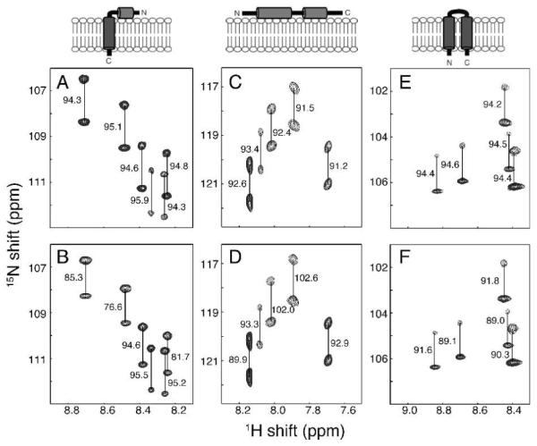
Representative regions of 1H-15N IPAP-HSQC spectra of isotropic (A, C and E) and aligned (B, D and F) samples. (A and B) Uniformly 15N labeled membrane-bound form of Pf1 coat protein in 100 mM DHPC, pH 6.7. (C and D) 15N-Ile labeled cytoplasmic domain of Vpu in 100 mM DHPC, 20 mM HEPES, pH 6.5. (E and F) Uniformly 15N-labeled MerFt in 100 mM DHPC, pH 6.5. The alignment resulted from the inclusion of 28 mg/ml, 13 mg/ml, and 30 mg/ml of fd bacteriophage in samples B, D, and F, respectively. The samples were maintained at 50°C for the measurements performed on a cryoprobe-equipped Bruker 600 MHz spectrometer. The measured values of the one-bond 1H-15N splittings are in Hz.
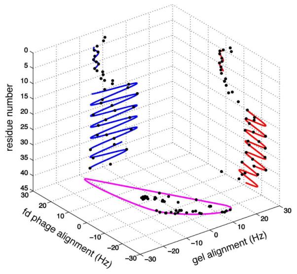
Cube correlation of Dipolar Waves and λ-map. Dipolar Waves fitted to the 1H-15N RDCs of membrane-bound form of Pf1 coat protein are shown for compressed polyacrylamide gel alignment (blue) and fd phage-induced alignment (red). λ-map estimate of the order tensors from both alignment media is shown in purple.
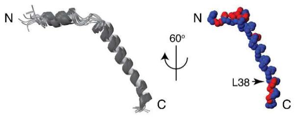
Superimposition of 15 calculated backbone structures of Pf1 coat protein (left) and 60° rotation of one structure to the vertical axis (right). A slight kink at residue 38 is indicated. Distribution of hydrophilic (red) and hydrophobic (blue) residues demonstrates the amphipathic character of the short N-terminal in-plane helix and the C-terminal region.
References
Publication types
MeSH terms
Substances
Grants and funding
LinkOut - more resources
Full Text Sources
