. 2009 Oct;10(10):1414-28.
doi: 10.1111/j.1600-0854.2009.00968.x.

Transportin mediates nuclear entry of DNA in vertebrate systems

Affiliations

Transportin mediates nuclear entry of DNA in vertebrate systems

Aurelie Lachish-Zalait et al. Traffic. 2009 Oct.

Abstract

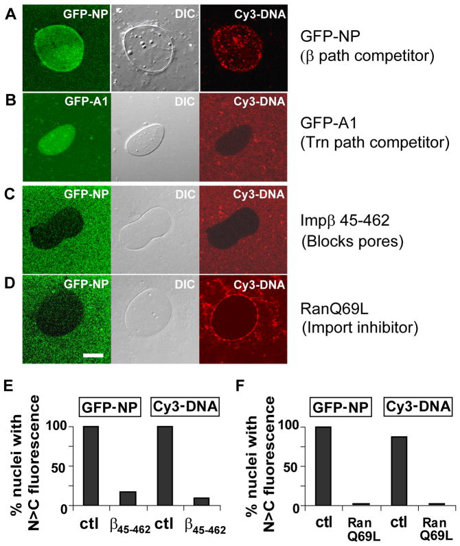
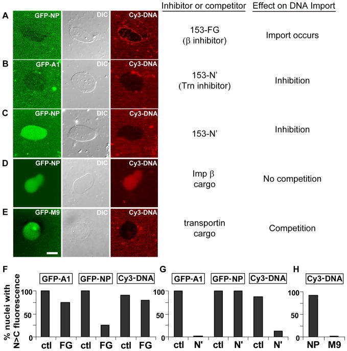
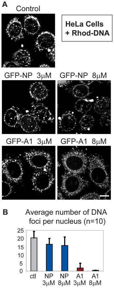
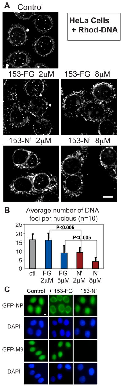
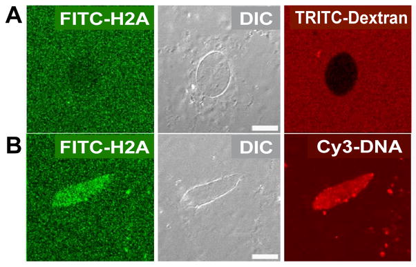
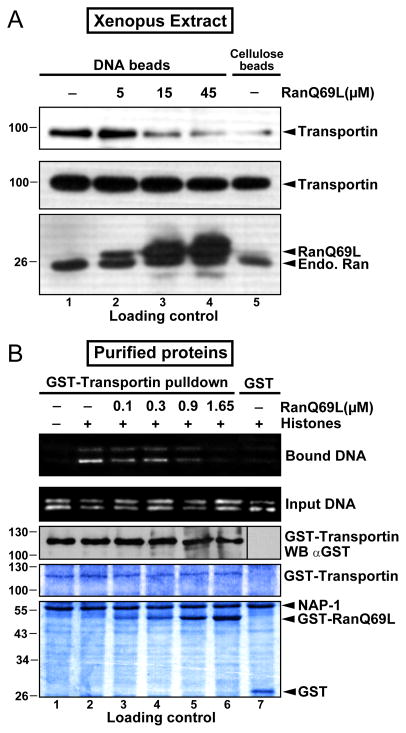
Delivery of DNA to the cell nucleus is an essential step in many types of viral infection, transfection, gene transfer by the plant pathogen Agrobacterium tumefaciens and in strategies for gene therapy. Thus, the mechanism by which DNA crosses the nuclear pore complex (NPC) is of great interest. Using nuclei reconstituted in vitro in Xenopus egg extracts, we previously studied DNA passage through the nuclear pores using a single-molecule approach based on optical tweezers. Fluorescently labeled DNA molecules were also seen to accumulate within nuclei. Here we find that this import of DNA relies on a soluble protein receptor of the importin family. To identify this receptor, we used different pathway-specific cargoes in competition studies as well as pathway-specific dominant negative inhibitors derived from the nucleoporin Nup153. We found that inhibition of the receptor transportin suppresses DNA import. In contrast, inhibition of importin beta has little effect on the nuclear accumulation of DNA. The dependence on transportin was fully confirmed in assays using permeabilized HeLa cells and a mammalian cell extract. We conclude that the nuclear import of DNA observed in these different vertebrate systems is largely mediated by the receptor transportin. We further report that histones, a known cargo of transportin, can act as an adaptor for the binding of transportin to DNA.


Figures

Figure 1
Figure 1. Scanning electron micrograph of a nucleus reconstituted in clarified crude egg extract
Numerous nuclear pores are observed at high magnification. Bars: 500 nm (large image) and 100 nm (inset).
Figure 2
Figure 2. DNA is imported into reconstituted nuclei, but is blocked by the global inhibitors importin β 45–462 and RanQ69L-GTP
Each row presents scanning confocal images of a representative nucleus in GFP, DIC, and Cy3 color channels. (A) Simultaneous accumulation of GFP-NP and Cy3-DNA is observed. (B) Nuclear accumulation of DNA is not observed in the presence of GFP-hnRNP A1. (C) Importin β 45–462 (FG-binding domain), which is a global inhibitor of all nuclear transport, inhibits GFP-NP and Cy3-DNA accumulation. (D) RanQ69L-GTP blocks nuclear uptake of both nucleoplasmin and DNA. (E–F) Bar graphs of replicate experiments for (C,D). The vertical axis denotes the relative percentage of nuclei scored positive for cargo accumulation. For assays involving soluble protein substrates, “positive” accumulation was scored if the nuclear concentration was higher than the cytoplasmic concentration. “Non-accumulation” was scored if equal or lower concentrations were found in the nucleus. DNA import was scored as positive for cases of either higher average nuclear intensity versus background, or for the appearance of intense nuclear DNA foci. Inhibitors and mock inhibition by addition of buffer (ctl) are listed on the horizontal axis. Nuclei were detected first by Hoechst staining and then checked for transport cargo in order to avoid biased data collection. The total number of nuclei counted for each treatment varied between 12 and 46. p-values obtained by Fisher’s exact test between ctl and β45–462 in (E) were p=0.000012 for GFP-NP and p<10−6 for Cy3-DNA. p-values between ctl and RanQ69L in (F) were p=0.0002 for GFP-NP and p=0.000002 for Cy3-DNA, thus all the p-values are significant. Bar: 10μm.
Figure 3
Figure 3. Multiple inhibitors of the transportin-mediated import pathway block DNA import
Each row shows scanning confocal images of a representative nucleus in GFP, DIC, and Cy3 color channels. (A) The importin β pathway inhibitor Nup153-FG effectively blocked the entry of nucleoplasmin. However, DNA uptake took place under these conditions, as observed from the punctate intranuclear foci that are typical of DNA import. (B) The transportin pathway inhibitor Nup153-N′ inhibited both GFP-A1 accumulation and DNA uptake. (C) Nup153-N′ fragment again inhibited DNA import, while nucleoplasmin accumulated in the nucleus. (D) DNA and nucleoplasmin, an importin β cargo, accumulated simultaneously in the nucleus, indicating a lack of competition between these substrates for transport factors. (E) GFP-M9, a transportin cargo, competed effectively with DNA, preventing its nuclear import. (F–H) Replicate measurements were scored and statistical tests performed as shown in Figure 2E–F. p-values between ctl and Nup153-FG in (F) were p=0.33 for GFP-A1, p=0.000147 for GFP-NP, and p=0.37 for Cy3-DNA import. This indicates significant inhibition by Nup153-FG of GFP-NP only. p-values between ctl and Nup153-N′ in (G) were p=0.008 for GFP-A1, p=1.000 for GFP-NP, and p=0.002 for Cy3-DNA import. This confirms significant inhibition by Nup153-N′ of GFP-A1 and Cy3-DNA, but not of GFP-NP. For (H), p=0.000003, confirming the suppression of DNA import by GFP-M9. Bar: 10 μm.
Figure 4
Figure 4. Excess transportin cargo, but not importin β cargo, blocks DNA import in permeabilized HeLa cells
CX-Rhodamine labeled DNA fragments of 400 bp were added to permeabilized HeLa cells in transport mixture, with or without competing protein substrates. Cells were incubated at 37°C for 90 minutes and fixed for confocal microscopy. (A) DNA imported into the nucleus was observed as distinct nuclear foci (control). DNA import was observed in the presence of excess of the importin α/β substrate, GFP-NP (GFP-NP, 3 or 8 μM), but not when excess GFP-A1, a transportin substrate, was present (GFP-A1, 3 or 8 μM). All images shown were projected from five z-sections at 0.5 μm spacing through the middle of the nuclei. Images were prepared using ImageJ software (http://rsb.info.nih.gov/ij/). (B) The number of nuclear DNA foci in 12 nuclei per condition was counted, the highest and lowest numbers were dropped, and the average and standard deviation were plotted. Bar: 10 μm.
Figure 5
Figure 5. A dominant negative inhibitor of the transportin pathway blocks DNA import in permeabilized HeLa cells
Permeabilized HeLa cell assays were performed and images were processed as in Figure 4. (A) In the presence of the importin β inhibitor Nup153-FG, a decrease in distinct nuclear foci of DNA was observed, but only at high concentration of inhibitor (153-FG, 8 μM). In the presence of the transportin inhibitor Nup153-N′, DNA import was suppressed at both high and low concentrations (153-N′, 2 and 8 μM). (B) The number of nuclear DNA foci was counted for each inhibitor. The average number of punctate nuclear foci per nucleus and the standard deviation are shown, as in Figure 4. p-values comparing DNA foci for 153-FG and 153-N′ inhibitors at the same concentrations (2 and 8 μM, respectively) were p<0.005, indicating a significant difference in their effectiveness. (C) In order to confirm specificity of the inhibition in this assay, Nup153-FG or Nup153-N′ was added at the final concentration of 25 μM, while GFP-NP or GFP-M9 was present at the final concentration of 3 μM or 4μM, respectively. Images shown are single planes through the z-axis. Bar: 10 μm.
Figure 6
Figure 6. Nuclear accumulation of histone H2A increases when exogenous DNA is imported
Fluorescein-histone H2A was added to reconstituted Xenopus nuclei in the absence or presence of 1500bp Cy3-DNA. In the absence of exogenous DNA, H2A gave a diffusion-like or weak exclusion pattern (A), while in the presence of 1500bp DNA, histone H2A clearly accumulated in nuclei (B) along with Cy3-DNA. Bars: 10 μm.
Figure 7
Figure 7. Core histones mediate interaction between transportin and DNA in vitro
(A) Xenopus laevis transportin interacts with DNA-conjugated cellulose beads and is removed by RanQ69L-GTP. High speed Xenopus laevis extract was pre-incubated with or without recombinant RanQ69L-GTP for 15 min on ice before the addition of DNA-conjugated cellulose beads and incubation at 4°C for 2 hr. After extensive washing, the DNA beads were treated with DNAse, and the bound proteins were eluted and processed for immunoblotting with anti-transportin or anti-Ran antibodies. In the top panel, lane 1 shows the amount of transportin bound to DNA cellulose in the absence of RanQ69L-GTP. Lanes 2–4 show the amount of transportin bound in the presence of the increasing concentrations of RanQ69L-GTP, and lane 5 shows the amount of transportin bound to cellulose beads lacking DNA. The bottom two panels are loading controls that show that equal amounts of transportin and endogenous Ran were present in each reaction, as determined by immunoblotting. (B) Drosophila melanogaster core histones mediate the interaction of transportin with DNA in a Ran-reversible manner. Recombinant GST-transportin was incubated with or without RanQ69L-GTP for 15 min on ice, before the addition of a mixture containing core histones, the histone chaperone NAP-1, and circular pEGFP-C2 plasmid DNA. Glutathione beads were then added and the reaction incubated overnight at 4°C, followed by six washes. The bound DNA was recovered (as described in Materials and Methods), separated on an agarose gel, and stained with Syber-green. For use as a loading control, 20% of the full reaction was removed before addition of the glutathione beads; half of this was treated with protease K to use for the DNA loading control (second panel; stained with Syber-green), while the other half was treated with DNAse to use for the protein loading controls. The proteins were separated on 10% SDS-PAGE and visualized with anti-GST immunoblotting (panel 3) or Coomassie blue (lower two panels).

Similar articles

Cited by

References

    1. Kasamatsu H, Nakanishi A. How do animal DNA viruses get to the nucleus? Annu Rev Microbiol. 1998;52:627–86. - PubMed
    1. Marsh M, Helenius A. Virus entry: open sesame. Cell. 2006;124:729–40. - PMC - PubMed
    1. Lechardeur D, Lukacs GL. Nucleocytoplasmic transport of plasmid DNA: a perilous journey from the cytoplasm to the nucleus. Hum Gene Ther. 2006;17:882–9. - PubMed
    1. Pouton CW, Wagstaff KM, Roth DM, Moseley GW, Jans DA. Targeted delivery to the nucleus. Adv Drug Deliv Rev. 2007;59:698–717. - PubMed
    1. Dowty ME, Williams P, Zhang G, Hagstrom JE, Wolff JA. Plasmid DNA entry into postmitotic nuclei of primary rat myotubes. Proc Natl Acad Sci U S A. 1995;92:4572–6. - PMC - PubMed

Publication types

LinkOut - more resources