. 2009 Sep 17:10:65.
doi: 10.1186/1471-2121-10-65.

Anti-proliferative activity of silver nanoparticles

Affiliations

Anti-proliferative activity of silver nanoparticles

P V Asharani et al. BMC Cell Biol. .

Abstract

Background: Nanoparticles possess exceptional physical and chemical properties which led to rapid commercialisation. Silver nanoparticles (Ag-np) are among the most commercialized nanoparticles due to their antimicrobial potential. Ag-np based cosmetics, therapeutic agents and household products are in wide use, which raised a public concern regarding their safety associated with human and environmental use. No safety regulations are in practice for the use of these nanomaterials. The interactions of nanomaterials with cells, uptake mechanisms, distribution, excretion, toxicological endpoints and mechanism of action remain unanswered.

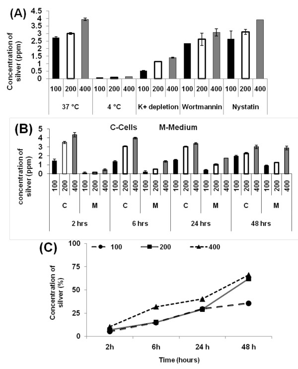
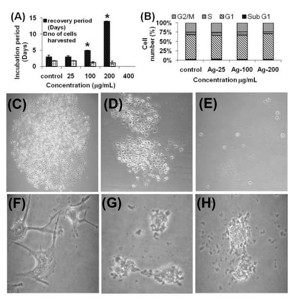
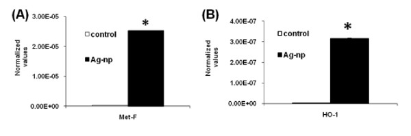
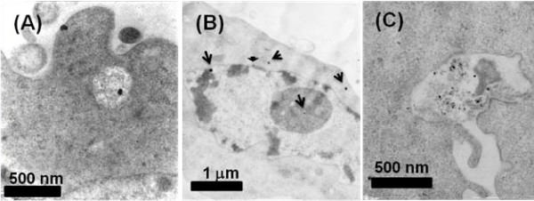
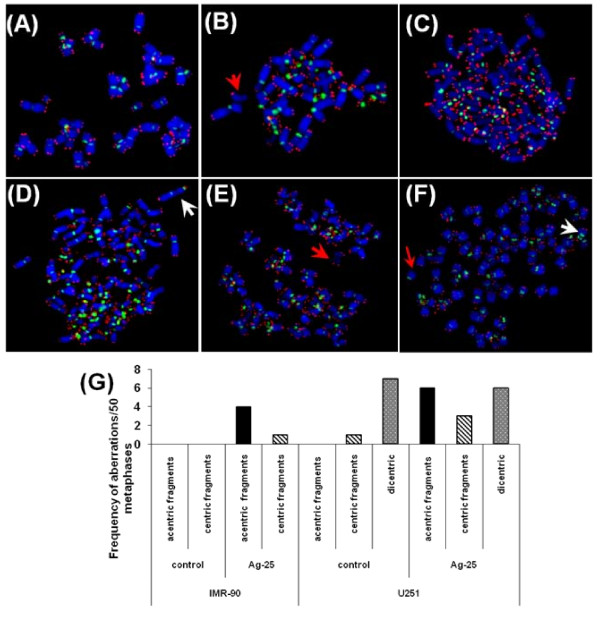
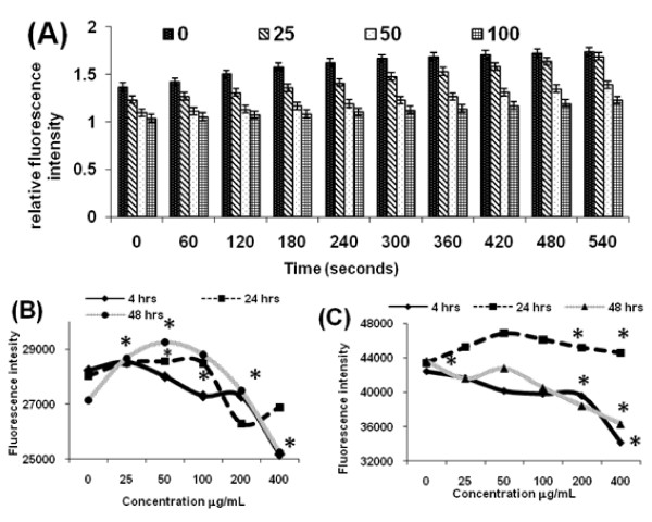
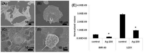
Results: Normal human lung fibroblasts (IMR-90) and human glioblastoma cells (U251) were exposed to different doses of Ag-nps in vitro. Uptake of Ag-nps occurred mainly through endocytosis (clathrin mediated process and macropinocytosis), accompanied by a time dependent increase in exocytosis rate. The electron micrographs revealed a uniform intracellular distribution of Ag-np both in cytoplasm and nucleus. Ag-np treated cells exhibited chromosome instability and mitotic arrest in human cells. There was efficient recovery from arrest in normal human fibroblasts whereas the cancer cells ceased to proliferate. Toxicity of Ag-np is mediated through intracellular calcium (Ca2+) transients along with significant alterations in cell morphology and spreading and surface ruffling. Down regulation of major actin binding protein, filamin was observed after Ag-np exposure. Ag-np induced stress resulted in the up regulation of metallothionein and heme oxygenase -1 genes.

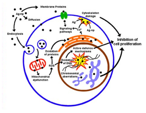
Conclusion: Here, we demonstrate that uptake of Ag-np occurs mainly through clathrin mediated endocytosis and macropinocytosis. Our results suggest that cancer cells are susceptible to damage with lack of recovery from Ag-np-induced stress. Ag-np is found to be acting through intracellular calcium transients and chromosomal aberrations, either directly or through activation of catabolic enzymes. The signalling cascades are believed to play key roles in cytoskeleton deformations and ultimately to inhibit cell proliferation.


Figures

Figure 1
Figure 1
Concentration of silver estimated using ICP-OES of Ag-np treated cells. (A) Data obtained from endocytosis studies using U251 cells. X axis represents different conditions employed viz. K+ depletion and temperature. Cells incubated at 37°C showed double the concentration of Ag than cells incubated at 4°C and K+ depleted environment. The concentration of Ag isolated from clathrin blocked cells and endocytosis blocked cells were approximately equal illustrating a clathrin independent process of uptake. (B) Exocytosis data show a concentration and time dependant exocytosis as indicated by the increased concentration of Ag in culture supernatant and a gradual decrease in cells. X axis represents different time and concentrations employed for exocytosis. Y axis represents concentration of silver detected by ICP-OES. (C) Comparison of exocytosis and endocytosis rate in U251 cells. Percentage of endocytosed nanoparticles expelled from cells over a period of time is depicted in the graph.
Figure 2
Figure 2
Recovery studies. Colony formation studies of Ag-np treated IMR-90 and U251 cells: The cells after Ag-np exposure were allowed to recover from stress. The time in days taken for the cells to reach confluence is expressed as the recovery period (Y-axis) (A). Concentrations of nanoparticles are indicated on the X- axis. Cells treated with all concentrations except 400 μg/mL recovered. *p < 0.05. (B) Cell cycle analysis of recovered cells showing absence of cell cycle arrest (n = 3). (C) Untreated cancer cells showing recovery and (D) Ag-np treated (25 μg/mL) cells forming colonies (1 week). (E). Cells treated with higher concentration of Ag-np (200 μg/mL) did not form colonies. The morphology of the cells under recovery deteriorated with time. (F) Control cells with proper protoplasmic extensions. (G) Ag-np treated cancer cells showing unhealthy cells with no proper protoplasmic extensions (2 weeks). (H) Morphological deterioration with time suggesting the onset of cell death cascades.
Figure 3
Figure 3
The RT-PCR profile. Expression profile of MT-1F (A) and HO-1 (B) in IMR-90 cells following Ag-np treatment. * p < 0.05 (n = 3). The values were normalised against the house keeping gene (18S RNA).
Figure 4
Figure 4
Electron micrographs of fibroblasts treated with Ag-np. TEM of Ag-np treated IMR-90 cells: Image shows presence of nanoparticles inside the phagosomes, near the cell membrane (A). Cell shows nanoparticles in the cytosol, nucleus and nucleoli. Arrow points to nanoparticles deposited. (B). Exocytic vesicles were observed at the cell periphery containing nanoparticles and cellular debris (C).
Figure 5
Figure 5
The chromosomal aberrations in IMR-90 and U251 cells. Metaphase spreads from the untreated cells show no apparent damage in the chromosomes (A), Ag-np treated IMR-90 cells show acentric and centric fragments (B). Arrow indicates acentric fragments. (C) Untreated cancer cells with no aberrations, metaphases show dicentric chromosomes in untreated cells (D) and treated cells. White arrow points to a dicentric chromosome. Cancer cells treated with Ag-np also show acentric fragments (E) and centric fragments (F). Red arrow points to a chromosome fragment. (G) Summary of the frequency of aberrations observed in Ag-np treated cells. A minimum of 50 metaphases per sample was scored for the chromosome analysis.
Figure 6
Figure 6
Calcium measurements. Calcium transients in Ag-np treated U251 and IMR-90 cells: Cells were stained with calcium binding fluorophore and subsequently activated with Ag-np (A). Data obtained from experiments where the U251 cells were treated with Ag-np for 4 hours and then stained with the dye (B). Graph denotes the calcium concentrations in Ag-np treated IMR-90 cells at 4, 24 and 48 hours. (C) U251 cells treated with Ag-np. * p < 0.05 (n = 4).
Figure 7
Figure 7
SEM images of fibroblasts and cancer cells: Untreated IMR-90 cells preserved their normal morphology (A). The Ag-np treated fibroblasts appeared more spherical and small with less cellular extensions (B). Untreated cancer cells showed normal morphology (C) whereas treated cells showed spherical cells with minimal cellular extensions and low spreading (D). (E) Filamin downregulation in normal and cancer cells as detected by RT-PCR.
Figure 8
Figure 8
The proposed mechanism of Ag-np toxicity based on the experimental data obtained in the present study.

References

    1. Percival SL, Bowler PG, Dolman J. Antimicrobial activity of silver-containing dressings on wound microorganisms using an in vitro biofilm model. Int Wound J. 2007;4:186–191. doi: 10.1111/j.1742-481X.2007.00296.x. - DOI - PMC - PubMed
    1. Yang P, Wei W, Tao C. Determination of trace thiocyanate with nano-silver coated multi-walled carbon nanotubes modified glassy carbon electrode. Anal Chim Acta. 2007;585:331–6. doi: 10.1016/j.aca.2007.01.011. - DOI - PubMed
    1. Kumar C. Nanomaterials- Toxicity, health and Environmental issues. Wiley- VCH Verlag GmbH & Co, Weinheim; 2006.
    1. Zhao Y, Nalwa H. Nanotoxicology - Interactions of Nanomaterials with Biological Systems. California, USA: American Scienfici Publishers; 2007.
    1. Elechiguerra JL, Burt JL, Morones JR, Camacho-Bragado A, Gao X, Lara HH, Yacaman MJ. Interaction of silver nanoparticles with HIV-1. J Nanobiotechnology. 2005;3:6. doi: 10.1186/1477-3155-3-6. - DOI - PMC - PubMed

Publication types

LinkOut - more resources