. 2010 Jan;156(Pt 1):116-127.
doi: 10.1099/mic.0.032318-0. Epub 2009 Sep 17.

Differential expression of Salmonella type III secretion system factors InvJ, PrgJ, SipC, SipD, SopA and SopB in cultures and in mice

Affiliations

Differential expression of Salmonella type III secretion system factors InvJ, PrgJ, SipC, SipD, SopA and SopB in cultures and in mice

Hao Gong et al. Microbiology (Reading). 2010 Jan.

Abstract

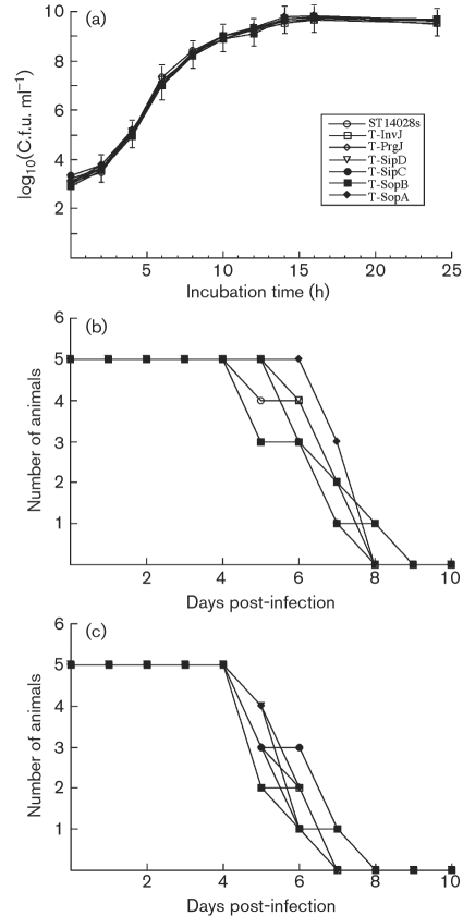
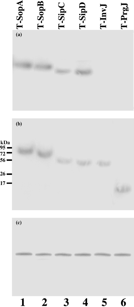
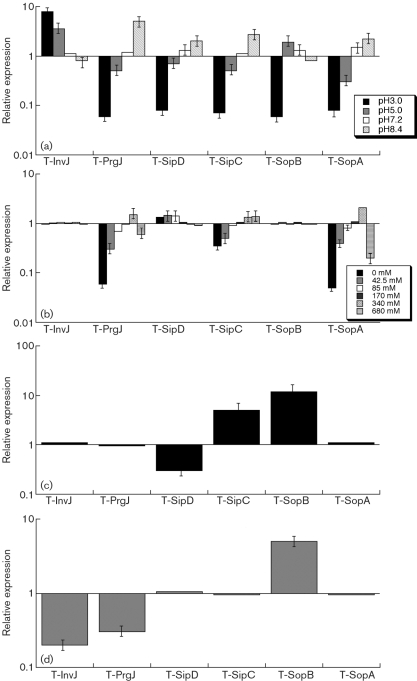
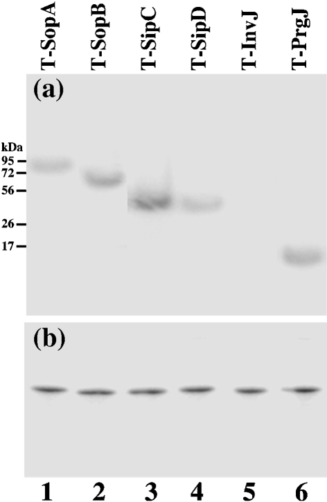
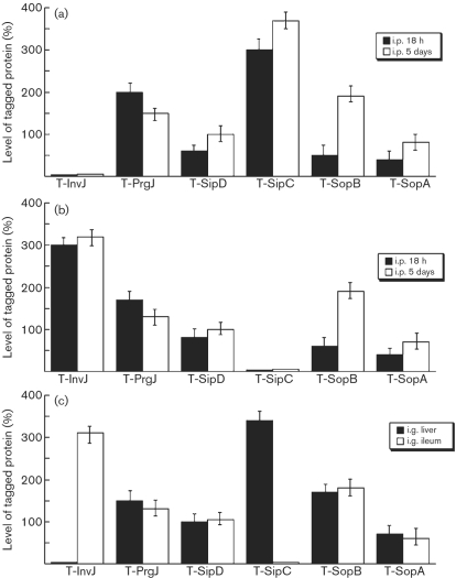
The type III secretion system (T3SS) encoded by Salmonella pathogenicity island 1 (SPI-1) is important for the invasion of epithelial cells during development of Salmonella-associated enterocolitis. It has been suggested that the level and timing of the expression of the SPI-1 T3SS proteins and effectors dictate the consequences of bacterial infection and pathogenesis. However, the expression of these proteins has not been extensively studied in vivo, especially during the later stages of salmonellosis when the infection is established. We have constructed recombinant Salmonella strains that contain a FLAG epitope inserted in-frame to genes invJ, prgJ, sipC, sipD, sopA and sopB, and investigated the expression of the tagged proteins both in vitro and in vivo during murine salmonellosis. Mice were inoculated intraperitoneally or intragastrically with the tagged Salmonella strains. At different time points post-infection, bacteria were recovered from various organs, and the expression of the tagged proteins was determined. Our results provide direct evidence that PrgJ and SipD are expressed in Salmonella colonizing the liver and ileum of infected animals at both the early and late stages of infection. Furthermore, our study has shown that the InvJ protein is expressed preferentially in Salmonella colonizing the ileum but not the liver, while SipC is expressed preferentially in Salmonella colonizing the liver but not the ileum. Thus, Salmonella appears to express different SPI-1 proteins and effectors when colonizing specific tissues. Our results suggest that differential expression of these proteins may be important for tissue-specific aspects of bacterial pathogenesis such as gastroenterititis in the ileum and systemic infection in the liver.


Figures

Fig. 1.
Fig. 1.
Growth analysis of bacterial strains in LB broth (a) and mortality of BALB/c (b) and SCID mice (c) infected with different strains. BALB/c mice (b) and CB17 SCID mice (c) (5 animals per group) were infected intragastrically with 5×10⁶ and 1×10³ c.f.u., respectively, of each bacterial strain. Means±sd are plotted.
Fig. 2.
Fig. 2.
Western blot analyses of the synthesis (b; cell extracts) and secretion (a; supernatants) of the tagged proteins from strains T-SopA (89 kDa) (lane 1), T-SopB (64 kDa) (lane 2), T-SipC (45 kDa) (lane 3), T-SipD (40 kDa) (lane 4), T-InvJ (39 kDa) (lane 5) and T-PrgJ (13 kDa) (lane 6). The expression of DnaK in bacterial cells was used as the internal control (c). Protein samples were separated in SDS-polyacrylamide gels and reacted with antibodies against the FLAG sequence (a, b) and DnaK (c). The molecular masses of some of the proteins in the PageRuler protein size markers (Fermentas) are shown. Each lane was loaded with lysates prepared from 5×10⁷ c.f.u. bacteria.
Fig. 3.
Fig. 3.
Effect of pH (a), osmolarity (b), limitation of oxygen (c), and the presence of butyrate (d) on the expression of the tagged proteins. The tagged strains were grown in culture media at different pH (a), at various concentrations of NaCl (b), in the presence and limitation of oxygen (c), or in the absence and presence of 10 mM butyrate (d) as described in Methods. The values of the relative expression, which are the means±sd from triplicate experiments, represent the ratios of the level of the tagged protein under the experimental conditions to the control pH 7.0 condition (a), to the control condition of 170 mM NaCl of regular LB broth (b), to the control condition of the presence of oxygen (c), or to the control condition of the absence of butyrate (d). The analyses were repeated at least three times.
Fig. 4.
Fig. 4.
Western blot analyses of the expression of the tagged proteins from strains T-SopA (lane 1), T-SopB (lane 2), T-SipC (lane 3), T-SipD (lane 4), T-InvJ (lane 5) and T-PrgJ (lane 6) recovered from the livers of infected mice. BALB/c mice were intraperitoneally infected with 1×10⁵ c.f.u. of the tagged strains, and bacteria were recovered from the livers at 5 days post-inoculation. Each lane was loaded with material from 5×10⁷ c.f.u. bacteria. Protein samples were reacted with antibodies against the FLAG sequence (a) and DnaK (b).
Fig. 5.
Fig. 5.
Level of tagged proteins from the bacterial strains recovered from the liver and ileum of infected mice. In (a) and (b), BALB/c mice were intraperitoneally (i.p.) infected with 1×10⁷ or 1×10⁵ c.f.u. of the tagged strains, and bacteria were recovered from the organs at 18 h or 5 days post-inoculation, respectively. The values, which are the means±sd from triplicate experiments, represent the relative percentage of the level of the tagged proteins in the bacteria recovered from the liver (a) and ileum (b), relative to the level of tagged SipD in strain T-SipD recovered from the liver and ileum, respectively, at 5 days post-inoculation (taken as 100 %). In (c), BALB/c mice were intragastrically (i.g.) infected with 1×10⁵ c.f.u. of the tagged strains, and bacteria were recovered from the liver and ileum at 7 days post-inoculation. The values, which are the means±sd from triplicate experiments, represent the level of the tagged proteins in the bacteria recovered from the organs relative to the level of tagged SipD in strain T-SipD recovered from the liver at 7 days post-inoculation.

References

    1. Abrahams, G. L. & Hensel, M. (2006). Manipulating cellular transport and immune responses: dynamic interactions between intracellular Salmonella enterica and its host cells. Cell Microbiol 8, 728–737. - PubMed
    1. Arricau, N., Hermant, D., Waxin, H., Ecobichon, C., Duffey, P. S. & Popoff, M. Y. (1998). The RcsB-RcsC regulatory system of Salmonella typhi differentially modulates the expression of invasion proteins, flagellin and Vi antigen in response to osmolarity. Mol Microbiol 29, 835–850. - PubMed
    1. Bajaj, V., Lucas, R. L., Hwang, C. & Lee, C. A. (1996). Co-ordinate regulation of Salmonella typhimurium invasion genes by environmental and regulatory factors is mediated by control of hilA expression. Mol Microbiol 22, 703–714. - PubMed
    1. Blanc-Potard, A. B., Solomon, F., Kayser, J. & Groisman, E. A. (1999). The SPI-3 pathogenicity island of Salmonella enterica. J Bacteriol 181, 998–1004. - PMC - PubMed
    1. Cain, R. J., Hayward, R. D. & Koronakis, V. (2008). Deciphering interplay between Salmonella invasion effectors. PLoS Pathog 4, e1000037. - PMC - PubMed

Publication types

LinkOut - more resources