

Distinct anatomical subtypes of the behavioural variant of frontotemporal dementia: a cluster analysis study
- PMID: 19762452
- PMCID: PMC2768663
- DOI: 10.1093/brain/awp232
Distinct anatomical subtypes of the behavioural variant of frontotemporal dementia: a cluster analysis study
Abstract
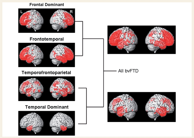
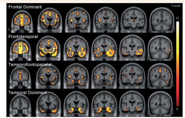
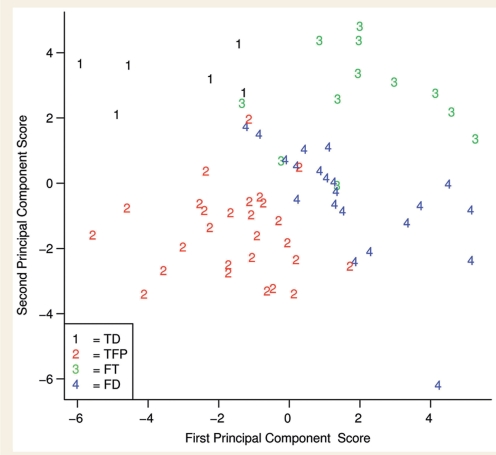
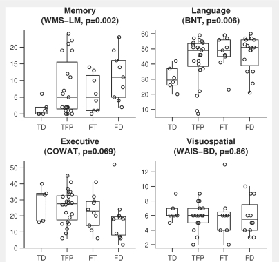
The behavioural variant of frontotemporal dementia is a progressive neurodegenerative syndrome characterized by changes in personality and behaviour. It is typically associated with frontal lobe atrophy, although patterns of atrophy are heterogeneous. The objective of this study was to examine case-by-case variability in patterns of grey matter atrophy in subjects with the behavioural variant of frontotemporal dementia and to investigate whether behavioural variant of frontotemporal dementia can be divided into distinct anatomical subtypes. Sixty-six subjects that fulfilled clinical criteria for a diagnosis of the behavioural variant of frontotemporal dementia with a volumetric magnetic resonance imaging scan were identified. Grey matter volumes were obtained for 26 regions of interest, covering frontal, temporal and parietal lobes, striatum, insula and supplemental motor area, using the automated anatomical labelling atlas. Regional volumes were divided by total grey matter volume. A hierarchical agglomerative cluster analysis using Ward's clustering linkage method was performed to cluster the behavioural variant of frontotemporal dementia subjects into different anatomical clusters. Voxel-based morphometry was used to assess patterns of grey matter loss in each identified cluster of subjects compared to an age and gender-matched control group at P < 0.05 (family-wise error corrected). We identified four potentially useful clusters with distinct patterns of grey matter loss, which we posit represent anatomical subtypes of the behavioural variant of frontotemporal dementia. Two of these subtypes were associated with temporal lobe volume loss, with one subtype showing loss restricted to temporal lobe regions (temporal-dominant subtype) and the other showing grey matter loss in the temporal lobes as well as frontal and parietal lobes (temporofrontoparietal subtype). Another two subtypes were characterized by a large amount of frontal lobe volume loss, with one subtype showing grey matter loss in the frontal lobes as well as loss of the temporal lobes (frontotemporal subtype) and the other subtype showing loss relatively restricted to the frontal lobes (frontal-dominant subtype). These four subtypes differed on clinical measures of executive function, episodic memory and confrontation naming. There were also associations between the four subtypes and genetic or pathological diagnoses which were obtained in 48% of the cohort. The clusters did not differ in behavioural severity as measured by the Neuropsychiatric Inventory; supporting the original classification of the behavioural variant of frontotemporal dementia in these subjects. Our findings suggest behavioural variant of frontotemporal dementia can therefore be subdivided into four different anatomical subtypes.
Figures


Comment in
-
With or without FUS, it is the anatomy that dictates the dementia phenotype.Brain. 2009 Nov;132(Pt 11):2906-8. doi: 10.1093/brain/awp286. Brain. 2009. PMID: 19861505 Free PMC article. No abstract available.
References
-
- Ashburner J, Friston KJ. Voxel-based morphometry—the methods. Neuroimage. 2000;11:805–21. - PubMed
-
- Ashburner J, Friston KJ. Unified segmentation. Neuroimage. 2005;26:839–51. - PubMed
-
- Baker M, Mackenzie IR, Pickering-Brown SM, Gass J, Rademakers R, Lindholm C, et al. Mutations in progranulin cause tau-negative frontotemporal dementia linked to chromosome 17. Nature. 2006;442:916–9. - PubMed
-
- Barnes J, Whitwell JL, Frost C, Josephs KA, Rossor M, Fox NC. Measurements of the amygdala and hippocampus in pathologically confirmed Alzheimer disease and frontotemporal lobar degeneration. Arch Neurol. 2006;63:1434–19. - PubMed
-
- Benton AL, Hamsher K. Manual. Iowa City: University of Iowa; 1989. Multilingual Aphasia Examination.
Publication types
MeSH terms
Substances
Grants and funding
LinkOut - more resources
Full Text Sources
Other Literature Sources
