

Case Reports
doi: 10.3174/ajnr.A1702.
Epub 2009 Sep 17.
Intra- and extracranial solitary fibrous tumor of the trigeminal nerve: CT and MR imaging appearance
Affiliations
- PMID: 19762459
- PMCID: PMC7964129
- DOI: 10.3174/ajnr.A1702
Item in Clipboard
Case Reports
Intra- and extracranial solitary fibrous tumor of the trigeminal nerve: CT and MR imaging appearance
AJNR Am J Neuroradiol.
2010 Feb.
Abstract
We describe a rare case of SFT existing along the mandibular division of the trigeminal nerve and extending down into the infratemporal fossa through the foramen ovale. The tumor showed heterogeneous hypointensity on T2-weighted images and marked enhancement on CT and MR images.
Figures
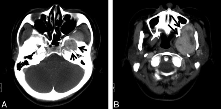
A, Axial contrast-enhanced CT scan shows an enlarged left foramen ovale (arrows) by the enhanced tumor. B, Axial contrast-enhanced CT scan of the caudal side of A shows the tumor compressing the maxillary bone, left pterygoid plate, and mandible (arrows), without destruction.
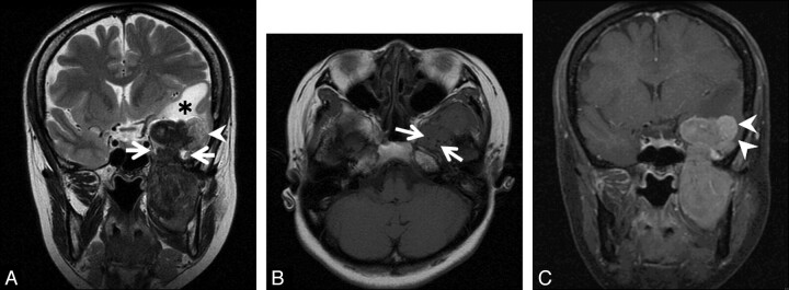
A, Coronal T2-weighted image shows a dumbbell-shaped tumor passing through left foramen ovale (arrows). The tumor is mainly hypointense to white matter. The lateral part of the intracranial component (arrowhead) shows higher intensities than other parts of the tumor. White matter of the left temporal lobe shows high intensities (asterisk). B, Axial T1-weighted image shows isointensity of the tumor to the brain. The tumor has low-intensity foci, suggesting a flow-void condition (arrows). C, On a coronal contrast-enhanced fat-suppressed T1-weighted image, the tumor shows heterogeneous enhancement. The lateral part of the intracranial component shows different strong enhancement (arrowheads).
References
-
- Carneiro SS, Scheithauer BW, Nascimento AG, et al. . Solitary fibrous tumor of the meninges: a lesion distinct from fibrous meningioma—a clinicopathologic and immunohistochemical study. Am J Clin Pathol 1996;106:217–24 - PubMed
-
- Bohinski RJ, Mendel E, Aldape KD, et al. . Intramedullary and extramedullary solitary fibrous tumor of the cervical spine: case report and review of the literature. J Neurosurg 2004;100:358–63 - PubMed
-
- Endo K, Komagata M, Ikegami H, et al. . Dumbbell-type solitary fibrous tumor in the cervical spine. J Orthop Sci 2003;8:428–31 - PubMed
-
- Kocak A, Cayli SR, Sarac K, et al. . Intraventricular solitary fibrous tumor: an unusual tumor with radiological, ultrastructural, and immunohistochemical evaluation—case report. Neurosurgery 2004;54:213–17 - PubMed
-
- Waldron JS, Tihan T, Parsa AT. Solitary fibrous tumor arising from cranial nerve VI in the prepontine cistern: case report and review of a tumor subpopulation mimicking schwannoma. Neurosurgery 2006;59:E939–40 - PubMed
Publication types
MeSH terms
LinkOut - more resources
Full Text Sources
Medical