

Intrathecal gadolinium-enhanced MR cisternography in the evaluation of CSF leakage
- PMID: 19762462
- PMCID: PMC7964096
- DOI: 10.3174/ajnr.A1788
Intrathecal gadolinium-enhanced MR cisternography in the evaluation of CSF leakage
Abstract
Background and purpose: Radiologic identification of the location of the CSF leakage is important for proper surgical planning and increases the chance of dural repair. This article describes our experience in analyzing clinically suspected cranial CSF fistulas by using MR imaging combined with the intrathecal administration of a gadolinium-based contrast agent.
Materials and methods: A total of 85 consecutive patients with suspected CSF fistulas who presented with persistent or intermittent rhinorrhea or otorrhea lasting for more than 1 month between 2003 and 2007 were included in this study.
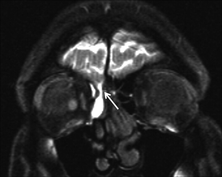
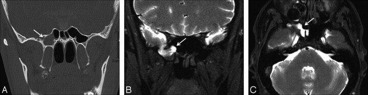
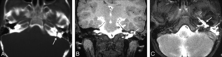
Results: We observed objective CSF leakage in 64 of 85 patients (75%). The CSF leak was located in the ethmoidal region in 37 patients (58%), in the superior wall of the sphenoid sinus in 8 patients (13%), in the posterior wall of the frontal sinus in 10 patients (15%), in the superior wall of the mastoid air cells in 6 patients (9%), and from the skull base into the infratemporal fossa in 1 patient (2%). Two patients (3%) showed leakage into >1 paranasal sinus.
Conclusions: MR cisternography after the intrathecal administration of gadopentate dimeglumine represents an effective and minimally invasive method for evaluating suspected CSF fistulas along the skull base. It provides multiplanar capabilities without risk of radiation exposure and is an excellent approach to depict the anatomy of CSF spaces and CSF fistulas.
Figures


References
-
- Park J, Strelzow V, Friedman W. Current management of cerebrospinal fluid rhinorrhea. Laryngoscope 1983; 93: 1924–300 - PubMed
-
- Johnson DB, Brennan P, Toland J, et al. . Magnetic resonance imaging in the evaluation of the cerebrospinal fluid fistula. Clin Radiol 1996; 51: 837–41 - PubMed
-
- Colquhoun IR. CT cisternography in the investigation of cerebrospinal rhinorrhea. Clin Radiol 1993; 47: 403–08 - PubMed
-
- Wakhloo AK, van Velthoven V, Shumacher M, et al. . Evaluation of MR imaging, digital subtraction cisternography, and CT cisternography in diagnosing CSF fistula. Acta Neurochir (Wien) 1991; 111: 119–27 - PubMed
-
- Manelfe C, Cellerier P, Sobel D, et al. . Cerebrospinal fluid rhinorrhea: evaluation with metrizamide cisternography. AJR Am J Roentgenol 1982; 138: 471–76 - PubMed
MeSH terms
Substances
LinkOut - more resources
Full Text Sources
Medical