. 2009 Nov 13;284(46):31616-24.
doi: 10.1074/jbc.M109.025882. Epub 2009 Sep 17.

Mitochondrial localization of PARP-1 requires interaction with mitofilin and is involved in the maintenance of mitochondrial DNA integrity

Affiliations

Mitochondrial localization of PARP-1 requires interaction with mitofilin and is involved in the maintenance of mitochondrial DNA integrity

Marianna N Rossi et al. J Biol Chem. .

Abstract

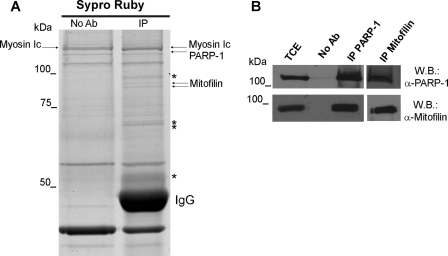
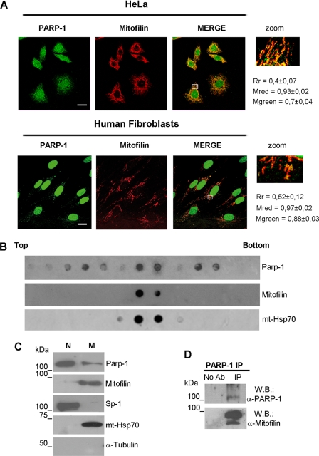
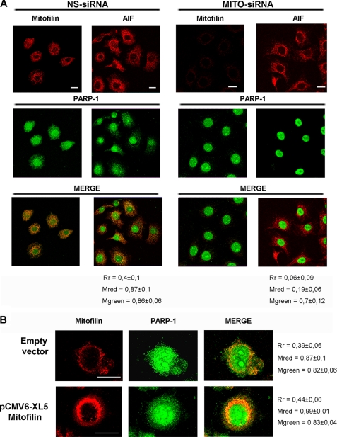
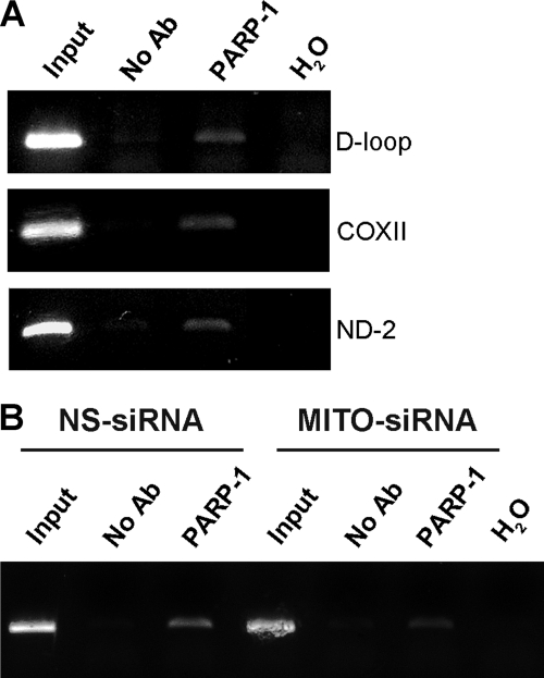
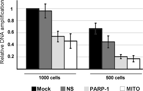
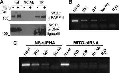
Poly(ADP-ribose)polymerase-1 (PARP-1) is a predominantly nuclear enzyme that exerts numerous functions in cellular physiology and pathology, from maintenance of DNA stability to transcriptional regulation. Through a proteomic analysis of PARP-1 co-immunoprecipitation complexes, we identified Mitofilin, a mitochondrial protein, as a new PARP-1 interactor. This result prompted us to further investigate the presence and the role of the enzyme in mitochondria. Using laser confocal microscopy and Western blot analysis of purified mitochondria, we demonstrated the mitochondrial localization of a fraction of PARP-1. Further, the effects of overexpressing or down-regulating Mitofilin showed that this protein promotes and is required for PARP-1 mitochondrial localization. We also report several lines of evidence suggesting that intramitochondrial PARP-1 plays a role in mitochondrial DNA (mtDNA) damage signaling and/or repair. First, we show that PARP-1 binds to different regions throughout the mtDNA. Moreover, we demonstrated that the depletion of either PARP-1 or Mitofilin, which abrogates the mitochondrial localization of the enzyme, leads to the accumulation of mtDNA damage. Finally, we show that DNA ligase III, known to be required for mtDNA repair, participates in a PARP-1-containing complex bound to mtDNA. This work highlights a new environment for PARP-1, opening the possibility that at least some of the nuclear functions of the enzyme can be also extended to mtDNA metabolism.


Figures

FIGURE 1.
FIGURE 1.
Interaction of PARP-1 with Mitofilin. A, PARP-1 immunoprecipitation was performed from human fibroblasts FB1329 using the monoclonal antibody F1-23 and SYPRO Ruby staining for proteomic analysis of co-immunoprecipitated proteins. Arrows indicate the protein identified by mass spectrometry. Bands marked with asterisks were identified as degradation products of IgG. No Ab, total cellular extracts incubated with protein A/G-Sepharose without antibody; IP, immunoprecipitation with the corresponding antibodies. B, PARP-1 and Mitofilin Western blots (W.B.) on PARP-1 and Mitofilin immunoprecipitates. TCE, total cellular extract.
FIGURE 2.
FIGURE 2.
PARP-1 interacts with Mitofilin within mitochondria. A, confocal laser scanning microscopy of double immunofluorescence staining performed on HeLa (upper panel) or FB 1329 (lower panel) cells. Fixed cells were labeled with α-PARP-1 (green) and α-Mitofilin (red). Bars indicate 20 μm. Rectangle-marked areas of each merge were enlarged in the zoom panels. Cytoplasmic co-localization was measured as described under “Experimental Procedures.” Rr, Pearson's correlation coefficient; Mred, Mander's co-localization coefficient for red; Mgreen, Mander's co-localization coefficient for green. B, mitochondria were purified from FB 1329 cells by a two-step sucrose gradient. The distribution of PARP-1, Mitofilin, and mtHsp70 in the gradient fractions was analyzed with the corresponding antibodies by dot blot. C, the nuclear fraction (N), obtained from differential centrifugation, and the mitochondrial fraction (M), obtained after sucrose gradient, were analyzed by Western blot with the listed antibodies. D, Mitofilin and PARP-1 Western blot (W.B.) on PARP-1 immunoprecipitates obtained from purified mitochondria. No Ab, mitochondrial extracts incubated with protein G-Sepharose without antibody; IP, immunoprecipitation with α-PARP-1 antibody.
FIGURE 3.
FIGURE 3.
Mitofilin is required for mitochondrial localization of PARP-1. A, HeLa cells were transfected with Mitofilin-specific (MITO-siRNA) or with nonspecific (NS-siRNA) siRNAs. After 72 h, cells were fixed for α-PARP-1/α-Mitofilin or α-PARP-1/α-AIF double immunofluorescence and confocal laser scanning analysis. AIF staining was shown as a mitochondrial marker in the absence of Mitofilin. B, HeLa cells were transfected with the pCMV6-XL5 vector containing the full-length human Mitofilin cDNA (OriGene) or with the empty vector. After 48 h, cells were fixed and labeled with α-PARP-1 and α-Mitofilin for confocal laser scanning microscopy. Bars indicate 20 μm. Cytoplasmic co-localization was measured as described under “Experimental Procedures.” Rr, Pearson's correlation coefficient; Mred, Mander's ccoefficient for red; Mgreen, Mander's co-localization coefficient for green.
FIGURE 4.
FIGURE 4.
Mitochondrial PARP-1 is associated to mitochondrial DNA. A, PCR analysis after chromatin immunoprecipitation with an antibody against PARP-1 showed the amplification of the desired product size in a different region of the mtDNA. DNA sample lane descriptions are as follows. Input, samples isolated from total lysates prior to antibody pulldown as an internal positive control; No Ab, samples isolated after pull down with no antibody; PARP-1, samples isolated after PARP-1 pull down; H2O, PCR negative control. B, HeLa cells were transfected with Mitofilin siRNA or control siRNA and analyzed as in A. The densitometric analysis of band intensity (ImageJ software) indicated that immunoprecipitation of Mitofilin-specific siRNA (MITO-siRNA)/nonspecific (NS-siRNA) siRNA = 0.5.
FIGURE 5.
FIGURE 5.
Mitochondrial PARP-1 is required for maintenance of DNA stability. HeLa cells were mock-transfected (Mock) or transfected with Mitofilin (MITO), PARP-1 (PARP-1), or nonspecific (NS) siRNAs and collected after 72 h for DPCR from whole cells. Two different amounts of cells (1000 and 500) were employed to ensure the reliability of the assay. Two pairs of primers were used to amplify two fragments of mtDNA corresponding, respectively, to a 157-bp region (used to normalize the mtDNA content) or to a 5-kbp region. The relative amplification efficiencies, calculated by densitometric analysis (ImageJ software) are shown as percentages of the control (Mock). The graph reports the mean values from three independent experiments.
FIGURE 6.
FIGURE 6.
Mitochondrial PARP-1 is associated with DNA Ligase III on mtDNA. A, PARP-1 was specifically immunoprecipitated from a mitochondria-enriched fraction of HeLa cells treated or not with 400 μm H2O2. Western blot (W.B.) analysis was performed with α-PARP-1 and α-DNA ligase III antibodies. mt, mitochondrial extract; No Ab, mitochondrial extracts incubated with protein G-Sepharose without antibody; IP, immunoprecipitation with α-PARP-1 antibody. B, sequential ChIP analysis of mtDNA from HeLa cells. Soluble chromatin was prepared from HeLa cells and divided into two chromatin aliquots, which were immunoprecipitated (first round of ChIP) with antibodies to PARP-1 and DNA ligase III, respectively. Immunocomplexes from PARP-1 and DNA ligase III were reimmunoprecipitated (second round of ChIP) with reciprocal antibodies (P/D and D/P). A fragment of the D-loop region was amplified by PCR. Input, samples isolated from total lysates prior to antibody pulldown as an internal positive control; No Ab, samples isolated after pulldown with no antibody; H2O, PCR negative control. C, sequential ChIP analysis of mtDNA from HeLa cell transfected with Mitofilin siRNA or control siRNA and analyzed as in B.

References

    1. Kim M. Y., Zhang T., Kraus W. L. (2005) Genes Dev. 19, 1951–1967 - PubMed
    1. D'Amours D., Desnoyers S., D'Silva I., Poirier G. G. (1999) Biochem. J. 342, 249–268 - PMC - PubMed
    1. Malanga M., Althaus F. R. (2005) Biochem. Cell Biol. 83, 354–364 - PubMed
    1. Saxena A., Saffery R., Wong L. H., Kalitsis P., Choo K. H. (2002) J. Biol. Chem. 277, 26921–26926 - PubMed
    1. Kraus W. L. (2008) Curr. Opin. Cell Biol. 20, 294–302 - PMC - PubMed

Publication types

MeSH terms