

Modulation of OPG, RANK and RANKL by human chondrocytes and their implication during osteoarthritis
- PMID: 19762475
- PMCID: PMC5250506
- DOI: 10.1093/rheumatology/kep300
Modulation of OPG, RANK and RANKL by human chondrocytes and their implication during osteoarthritis
Abstract
Objectives: Earlier studies suggest the involvement of osteoprotegerin (OPG), RANK and RANK ligand (RANKL) in OA subchondral bone metabolism; however, few studies have looked at their functional consequences on chondrocytes. We compared the expression/production of OPG, RANK and RANKL on human normal and OA chondrocytes, and evaluated, on OA chondrocytes, their modulation by some catabolic factors. Furthermore, the role of OPG and RANKL on the production of catabolic/anabolic factors was assessed.
Methods: Expression was determined using real-time PCR, production of RANK and RANKL by flow cytometry and that of OPG by ELISA. Modulation of these factors was determined upon treatment with IL-1beta, TNF-alpha and PGE(2). The functional consequences were examined following treatment with soluble RANKL or OPG-Fc (OPG without the heparin-binding domain).
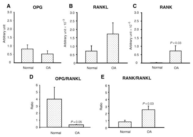
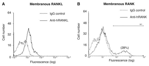
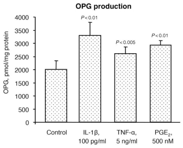
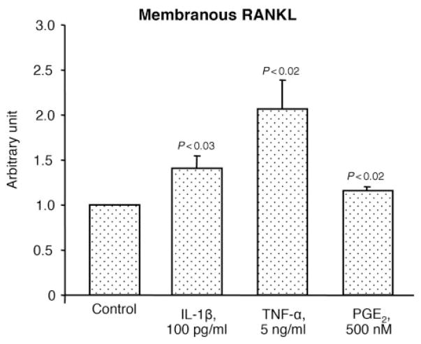
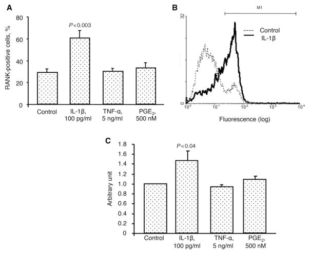
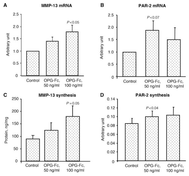
Results: OPG, RANK and RANKL were expressed and produced by human chondrocytes. Membranous RANK was produced only by an OA chondrocyte subpopulation (29%) localized throughout the cartilage. The OPG/RANKL ratio was significantly (P = 0.05) reduced on the OA chondrocytes, whereas the RANK/RANKL ratio was significantly (P < 0.03) increased. OPG and membranous RANKL levels were significantly enhanced by IL-1beta, TNF-alpha and PGE(2), whereas membranous RANK was significantly increased only with IL-1beta. Administration of soluble RANKL had no effect on the OA chondrocytes. However, addition of OPG-Fc significantly stimulated MMP-13 (P = 0.05) and protease-activated receptor-2 (PAR-2) (P < 0.04) production.
Conclusions: Our findings showed that human chondrocytes express and produce OPG, RANK and RANKL. OA chondrocyte treatment with catabolic factors pointed towards an increased biological effect of OPG. Interestingly, OPG appears to be involved in OA progression by increasing two catabolic factors involved in cartilage pathophysiology.
Conflict of interest statement
statement: The authors have declared no conflicts of interest.
Figures
References
-
- Radin EL, Rose RM. Role of subchondral bone in the initiation and progression of cartilage damage. Clin Orthop. 1986;213:34–40. - PubMed
-
- Bailey AJ, Mansell JP. Do subchondral bone changes exacerbate or precede articular cartilage destruction in osteoarthritis of the elderly? Gerontology. 1997;43:296–304. - PubMed
-
- Mansell JP, Tarlton JF, Bailey AJ. Biochemical evidence for altered subchondral bone collagen metabolism in osteoarthritis of the hip. Br J Rheumatol. 1997;36:16–19. - PubMed
-
- Lajeunesse D, Massicotte F, Pelletier JP, Martel-Pelletier J. Subchondral bone sclerosis in osteoarthritis: not just an innocent bystander. Mod Rheumatol. 2003;13:7–14. - PubMed
-
- Martel-Pelletier J, Lajeunesse D, Pelletier JP. Etiopathogenesis of osteoarthritis. In: Koopman Moreland., editor. Arthritis & allied conditions. A textbook of rheumatology. 15. Baltimore: Lippincott, Williams & Wilkins; 2005. pp. 2199–226.
